Division of Hypertension and Vascular Research, Henry Ford Health System, Detroit, MI, USA.
Division of Hypertension and Vascular Research, Henry Ford Health System, Detroit, MI, USA.
Biochem Biophys Res Commun. 2019 May 21;513(1):166-171. doi: 10.1016/j.bbrc.2019.03.177. Epub 2019 Apr 2.
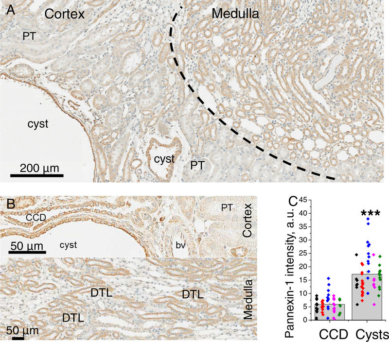
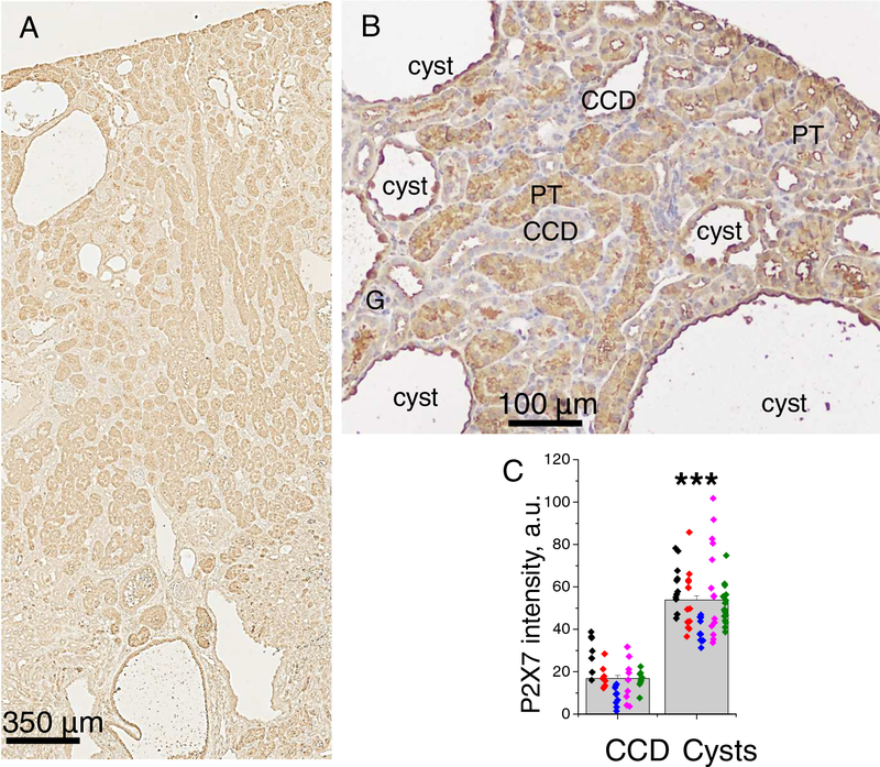
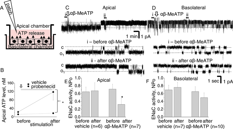
Genetic predisposition is necessary for polycystic kidney disease (PKD) initiation, although there are other, incompletely identified downstream processes that are required for cyst growth. Their characterization may provide a unique opportunity for clinical interventions. One of the poorly studied phenomena in PKD is high ATP content in cysts. Unfortunately, neither origins of uncontrolled ATP release, nor consequences of abnormal purinergic signaling in relation to epithelial transport are well explored in the polycystic kidney. We tested the distribution of pannexin-1 (Panx1) and P2X7, two proteins potentially involved in ATP release, in the kidneys of the Pkd1 mice, a model of autosomal dominant PKD (ADPKD). Abundances of both proteins were abnormally increased in the cyst lining cells compared to non-dilated collecting ducts. To establish if pannexin-1 contributes to ATP release in the collecting ducts (CD), we measured luminal accumulation of ATP in M1 cell renal CD monolayers, and found that treatment with probenecid, a Panx1 blocker, prevents ATP release. Single channel patch clamp analysis of polarized M1 cells revealed that apical stimulation of P2X receptors with αβ-MeATP acutely reduces ENaC activity. We conclude that in ADPKD progression, an abnormal hyperexpression of both PANX1 and P2RX7 occurs in the cyst lining epithelial cells. High abundance of both proteins is not typical for non-dilated CDs but, when it happens in cysts, pannexin1/P2X7 cooperation elevates ATP release into the luminal space. High ATP level is a pathogenic factor facilitating cystogenesis by reducing ENaC-mediated reabsorption from the lumen.
遗传易感性是多囊肾病 (PKD) 发生的必要条件,尽管还存在其他不完全确定的下游过程,这些过程对于囊肿生长是必需的。对这些过程的特征进行描述可能为临床干预提供独特的机会。在 PKD 中研究不足的现象之一是囊肿中存在高含量的 ATP。不幸的是,在多囊肾中,尚未对不受控制的 ATP 释放的起源,或与上皮转运有关的异常嘌呤能信号传导的后果进行深入研究。我们检测了 Panx1 和 P2X7 两种可能参与 ATP 释放的蛋白在 Pkd1 小鼠(一种常染色体显性多囊肾病的模型)肾脏中的分布。与非扩张的集合管相比,这两种蛋白在囊泡衬里细胞中的丰度异常增加。为了确定 Panx1 是否有助于集合管 (CD) 中的 ATP 释放,我们测量了 M1 细胞肾 CD 单层细胞腔中的 ATP 积累,发现 Panx1 阻断剂丙磺舒处理可防止 ATP 释放。极化 M1 细胞的单通道膜片钳分析表明,用 αβ-MeATP 刺激顶端的 P2X 受体可急性降低 ENaC 活性。我们的结论是,在 ADPKD 进展过程中,囊泡衬里上皮细胞中异常过度表达了 PANX1 和 P2RX7。这两种蛋白的高丰度并不典型,但当它们出现在囊肿中时,pannexin1/P2X7 合作会增加 ATP 向腔空间的释放。高水平的 ATP 是一种促病变因子,通过减少从腔内向细胞的 ENaC 介导的重吸收,促进囊肿形成。