Department of Pharmaceutical Sciences, School of Pharmacy, University of Pittsburgh, Pittsburgh, Pennsylvania.
Department of Computational and Systems Biology, School of Medicine, University of Pittsburgh, Pittsburgh, Pennsylvania.
Cancer Res. 2019 Jun 1;79(11):2962-2977. doi: 10.1158/0008-5472.CAN-18-3151. Epub 2019 Apr 5.
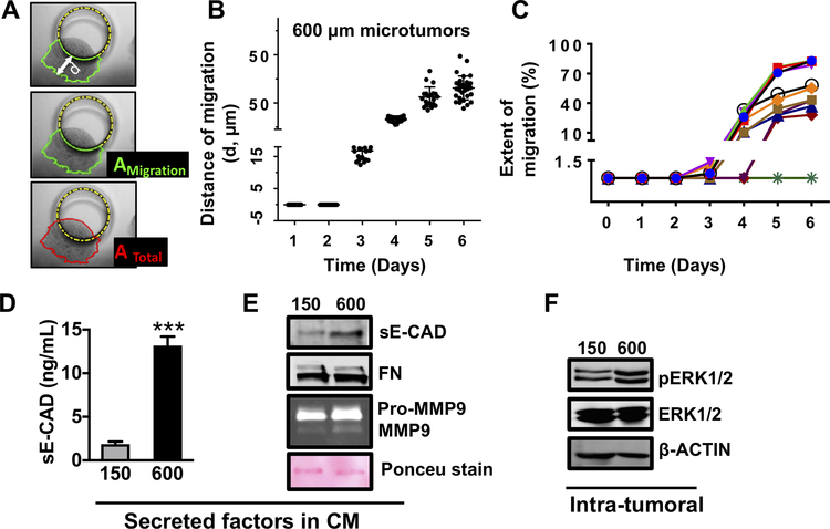
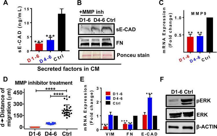
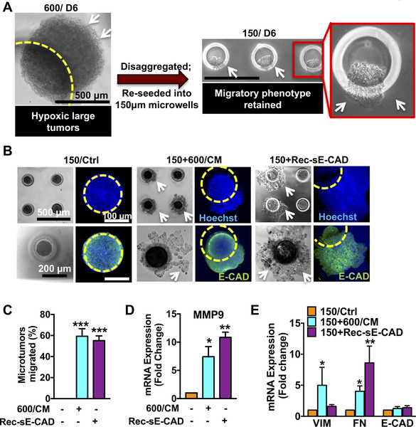
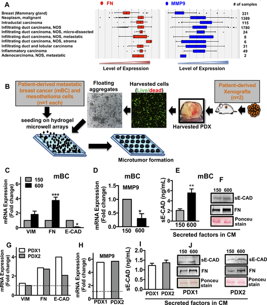
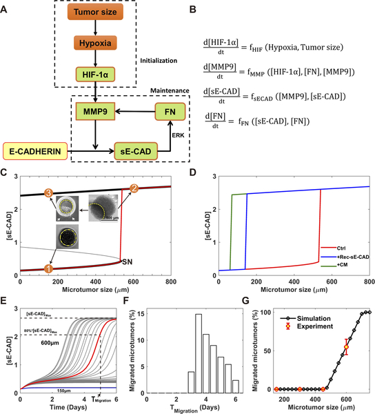
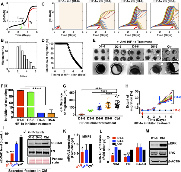
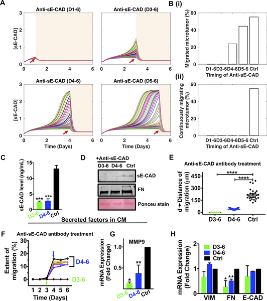
Targeting microenvironmental factors that foster migratory cell phenotypes is a promising strategy for halting tumor migration. However, lack of mechanistic understanding of the emergence of migratory phenotypes impedes pharmaceutical drug development. Using our three-dimensional microtumor model with tight control over tumor size, we recapitulated the tumor size-induced hypoxic microenvironment and emergence of migratory phenotypes in microtumors from epithelial breast cells and patient-derived primary metastatic breast cancer cells, mesothelioma cells, and lung cancer xenograft cells. The microtumor models from various patient-derived tumor cells and patient-derived xenograft cells revealed upregulation of tumor-secreted factors, including matrix metalloproteinase-9 (MMP9), fibronectin (FN), and soluble E-cadherin, consistent with clinically reported elevated levels of FN and MMP9 in patient breast tumors compared with healthy mammary glands. Secreted factors in the conditioned media of large microtumors induced a migratory phenotype in nonhypoxic, nonmigratory small microtumors. Subsequent mathematical analyses identified a two-stage microtumor progression and migration mechanism whereby hypoxia induces a migratory phenotype in the initialization stage, which then becomes self-sustained through a positive feedback loop established among the tumor-secreted factors. Computational and experimental studies showed that inhibition of tumor-secreted factors effectively halts microtumor migration despite tumor-to-tumor variation in migration kinetics, while inhibition of hypoxia is effective only within a time window and is compromised by tumor-to-tumor variation, supporting our notion that hypoxia initiates migratory phenotypes but does not sustain it. In summary, we show that targeting temporal dynamics of evolving microenvironments, especially tumor-secreted factors during tumor progression, can halt tumor migration. SIGNIFICANCE: This study uses state-of-the-art three-dimensional microtumor models and computational approaches to highlight the temporal dynamics of tumor-secreted microenvironmental factors in inducing tumor migration.
靶向促进迁移细胞表型的微环境因素是阻止肿瘤迁移的一种有前途的策略。然而,对迁移表型出现的机制理解不足阻碍了药物的开发。我们使用对肿瘤大小具有严格控制的三维微肿瘤模型,重现了大小诱导的缺氧微环境以及上皮性乳腺癌细胞和源自患者的原发性转移性乳腺癌细胞、间皮瘤细胞和肺癌异种移植细胞的微肿瘤中迁移表型的出现。来自各种源自患者的肿瘤细胞和源自患者的异种移植细胞的微肿瘤模型显示,肿瘤分泌因子(包括基质金属蛋白酶 9(MMP9)、纤维连接蛋白(FN)和可溶性 E-钙黏蛋白)上调,与临床报道的患者乳腺肿瘤中 FN 和 MMP9 水平升高一致。大微肿瘤条件培养基中的分泌因子诱导非缺氧、非迁移的小微肿瘤出现迁移表型。随后的数学分析确定了微肿瘤进展和迁移的两阶段机制,其中缺氧在初始化阶段诱导迁移表型,然后通过肿瘤分泌因子之间建立的正反馈回路维持自身。计算和实验研究表明,尽管肿瘤间在迁移动力学上存在差异,但抑制肿瘤分泌因子可有效阻止微肿瘤迁移,而抑制缺氧仅在一定时间窗口内有效,并且受到肿瘤间差异的影响,这支持了我们的观点,即缺氧引发迁移表型,但不能维持它。总之,我们表明靶向不断发展的微环境的时间动态,特别是肿瘤进展过程中的肿瘤分泌因子,可以阻止肿瘤迁移。意义:本研究使用最先进的三维微肿瘤模型和计算方法,强调了肿瘤分泌的微环境因子在诱导肿瘤迁移中的时间动态。