Department of Oncology, CRUK/MRC Oxford Institute for Radiation Oncology, University of Oxford, Oxford, United Kingdom.
Proteases and Tissue Remodeling Section, National Institute of Dental and Craniofacial Research, National Institutes of Health, Bethesda, Maryland.
J Nucl Med. 2019 Oct;60(10):1474-1482. doi: 10.2967/jnumed.119.226423. Epub 2019 Apr 6.
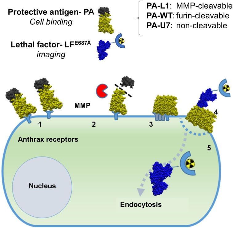
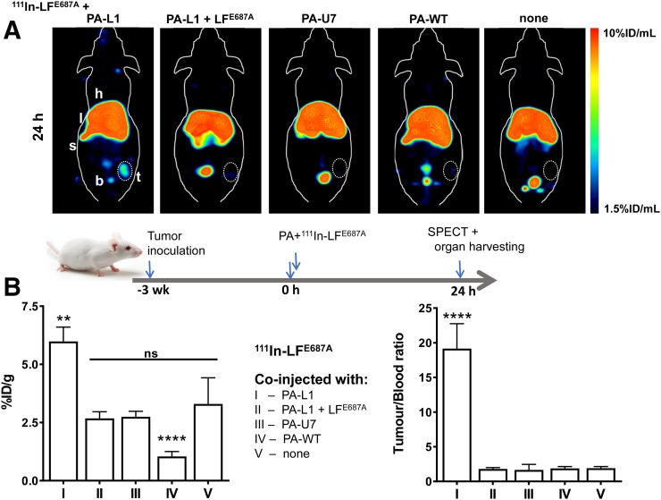
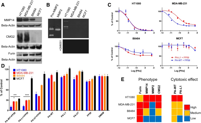
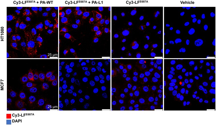
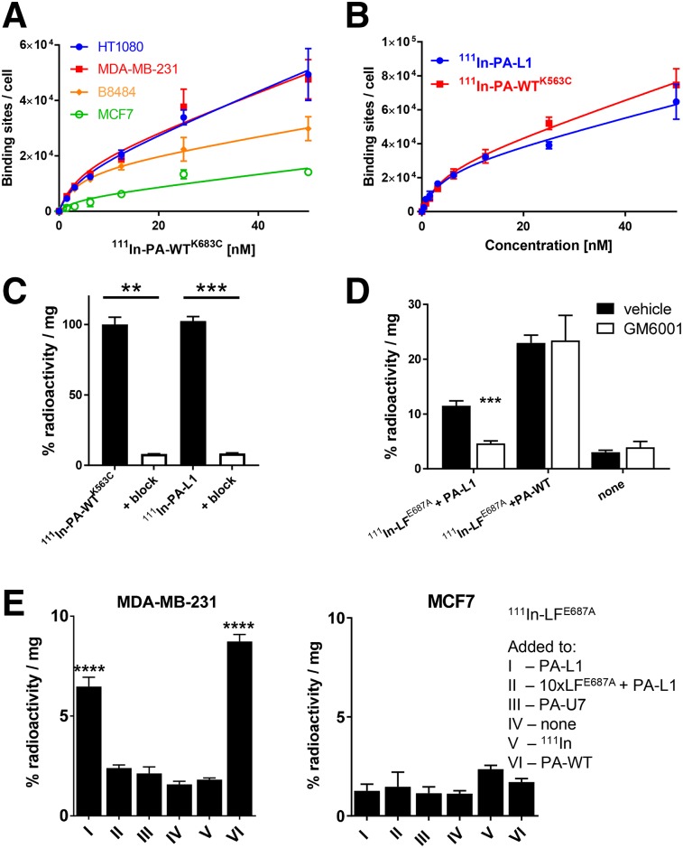
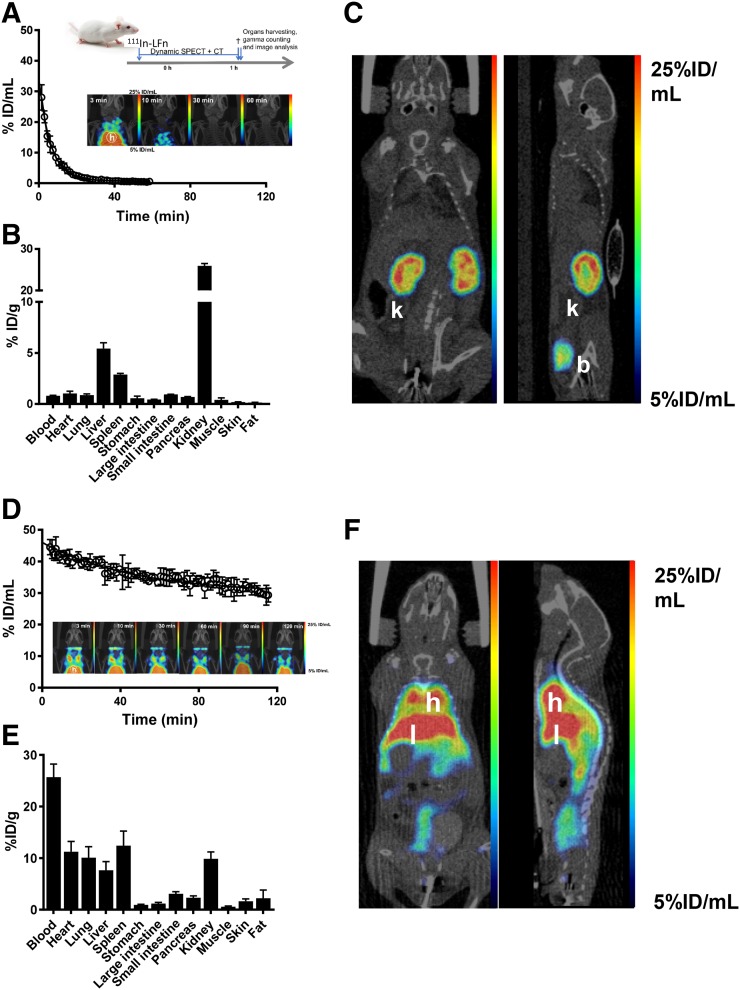
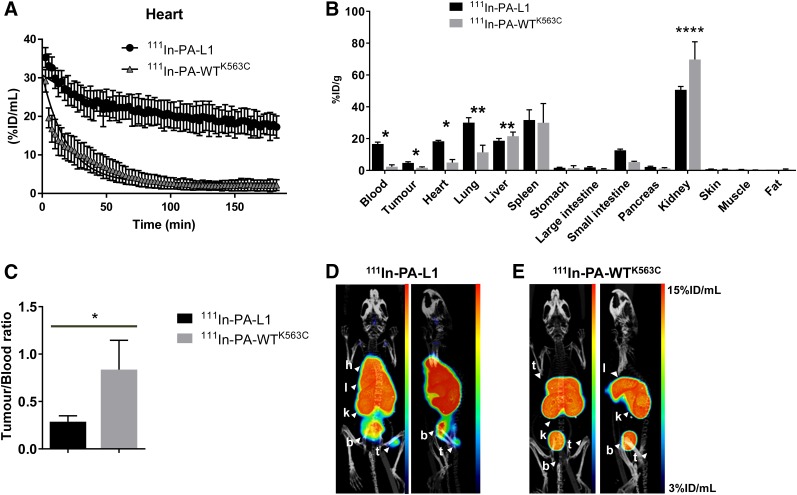
Increased activity of matrix metalloproteinases (MMPs) is associated with worse prognosis in different cancer types. The wild-type protective antigen (PA-WT) of the binary anthrax lethal toxin was modified to form a pore in cell membranes only when cleaved by MMPs (to form PA-L1). Anthrax lethal factor (LF) is then able to translocate through these pores. Here, we used a In-radiolabeled form of LF with the PA/LF system for noninvasive in vivo imaging of MMP activity in tumor tissue by SPECT. MMP-mediated activation of PA-L1 was correlated to anthrax receptor expression and MMP activity in a panel of cancer cells (HT1080, MDA-MB-231, B8484, and MCF7). Uptake of In-radiolabeled PA-L1, In-PA-WT, or In-LF (a catalytically inactive LF mutant) in tumor and normal tissues was measured using SPECT/CT imaging in vivo. Activation of PA-L1 in vitro correlated with anthrax receptor expression and MMP activity (HT1080 > MDA-MB-231 > B8484 > MCF7). PA-L1-mediated delivery of In-LF was demonstrated and was corroborated using confocal microscopy with fluorescently labeled LF Uptake was blocked by the broad-spectrum MMP inhibitor GM6001. In vivo imaging showed selective accumulation of In-PA-L1 in MDA-MB-231 tumor xenografts (5.7 ± 0.9 percentage injected dose [%ID]/g) at 3 h after intravenous administration. In-LF was selectively delivered to MMP-positive MDA-MB-231 tumor tissue by MMP-activatable PA-L1 (5.98 ± 0.62 %ID/g) but not by furin-cleavable PA-WT (1.05 ± 0.21 %ID/g) or a noncleavable PA variant control, PA-U7 (2.74 ± 0.24 %ID/g). Taken together, our results indicate that radiolabeled forms of mutated anthrax lethal toxin hold promise for noninvasive imaging of MMP activity in tumor tissue.
基质金属蛋白酶(MMPs)活性增加与不同癌症类型的预后不良有关。二元炭疽致死毒素的野生型保护抗原(PA-WT)经过修饰,只有在被 MMP 切割时才能在细胞膜上形成孔(形成 PA-L1)。炭疽致死因子(LF)随后能够通过这些孔转移。在这里,我们使用一种放射性标记的 LF 形式与 PA/LF 系统一起,通过 SPECT 对肿瘤组织中的 MMP 活性进行非侵入性体内成像。MMP 介导的 PA-L1 激活与炭疽受体表达和一系列癌细胞(HT1080、MDA-MB-231、B8484 和 MCF7)中的 MMP 活性相关。使用 SPECT/CT 成像在体内测量肿瘤和正常组织中放射性标记的 PA-L1、In-PA-WT 或 In-LF(一种催化失活的 LF 突变体)的摄取。体外 PA-L1 的激活与炭疽受体表达和 MMP 活性相关(HT1080 > MDA-MB-231 > B8484 > MCF7)。已经证明了 PA-L1 介导的 In-LF 的递送,并使用带有荧光标记 LF 的共聚焦显微镜进行了证实。摄取被广谱 MMP 抑制剂 GM6001 阻断。体内成像显示,静脉注射后 3 小时,In-PA-L1 在 MDA-MB-231 肿瘤异种移植中选择性积累(5.7 ± 0.9 注射剂量百分比 [%ID]/g)。MMP 激活的 PA-L1 (5.98 ± 0.62 %ID/g)而非 furin 切割的 PA-WT (1.05 ± 0.21 %ID/g)或非切割的 PA 变体对照 PA-U7 (2.74 ± 0.24 %ID/g)将 In-LF 选择性递送至 MMP 阳性 MDA-MB-231 肿瘤组织。总之,我们的结果表明,放射性标记的突变炭疽致死毒素形式有望用于肿瘤组织中 MMP 活性的非侵入性成像。