Gharbaran Rajendra, Zhang Bo, Valerio Luis, Onwumere Onyekwere, Wong Madeline, Mighty Jason, Redenti Stephen
Department of Biological Sciences, Bronx Community College, The City University of New York, Bronx, NY, 10453, USA.
Department of Biological Sciences, Lehman College, City University of New York, Bronx, NY, 10468, USA.
BMC Res Notes. 2019 Apr 8;12(1):216. doi: 10.1186/s13104-019-4241-0.
Vitamin D receptor (VDR) activities have been noted for a number of B cell malignancies which showed varying sensitivities to vitamin D3 (1,25-dihydroxyvitamin D3, VD3, calcitriol) and its synthetic analogs. The objective of this study was to address the potential effects of VD3 and vitamin D3 analogs (VDAs) on the growth of Hodgkin's lymphoma (HL), a malignant pathology of B cell origin, in vitro.
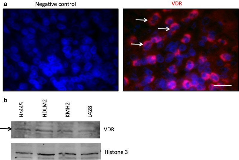
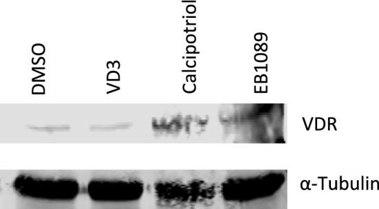
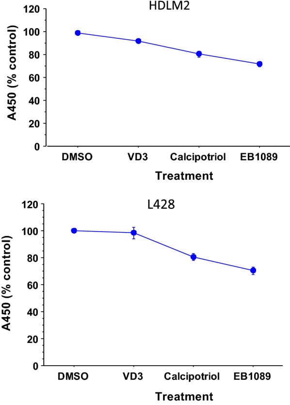
Immunofluorescence staining showed the expression of VDR by primary Hodgkin's (H) and Reed-Sternberg (RS)-HRS-tumor cells in HL histological sections. Western blot analyses revealed expression of VDR in the HL cell lines Hs445, HDLM2, KMH2, and L428. One-way analysis of variance (ANOVA) on data obtained from water-soluble tetrazolium 1 (WST-1) cell proliferation assay showed decreased cell growth in HDLM2 and L428, 72 h after treatment with 10 µM of either VD3 of VDAs. Western blot analyses showed that treatment of L428 cells with the VDAs (calcipotriol and EB1089) resulted in modest increases in nuclear accumulation of VDR (nuVDR) compared to either dimethyl sulfoxide (DMSO) or VD3 treatments. nuVDR for DMSO control and VD3 was comparable. These results suggest that VD3 or VDAs may affect growth of HL.
已注意到维生素D受体(VDR)活性在多种B细胞恶性肿瘤中存在,这些肿瘤对维生素D3(1,25 - 二羟维生素D3,VD3,骨化三醇)及其合成类似物表现出不同的敏感性。本研究的目的是探讨VD3和维生素D3类似物(VDAs)对霍奇金淋巴瘤(HL)(一种B细胞起源的恶性病变)体外生长的潜在影响。
免疫荧光染色显示HL组织切片中原发性霍奇金(H)和里德 - 斯腾伯格(RS) - HRS肿瘤细胞表达VDR。蛋白质印迹分析显示HL细胞系Hs445、HDLM2、KMH2和L428中存在VDR表达。对水溶性四氮唑盐1(WST - 1)细胞增殖试验获得的数据进行单因素方差分析(ANOVA)表明,用10 μM的VD3或VDAs处理72小时后,HDLM2和L428中的细胞生长减少。蛋白质印迹分析表明,与二甲基亚砜(DMSO)或VD3处理相比,用VDAs(卡泊三醇和EB1089)处理L428细胞导致VDR核积累(nuVDR)适度增加。DMSO对照和VD3的nuVDR相当。这些结果表明VD3或VDAs可能影响HL的生长。