Department of Cardiology, University of Heidelberg, 69120 Heidelberg, Germany.
DZHK (German Center for Cardiovascular Research), Partner Site Heidelberg/Mannheim, University of Heidelberg, 69120 Heidelberg, Germany.
Mol Biol Cell. 2019 Jun 1;30(12):1425-1436. doi: 10.1091/mbc.E18-10-0687. Epub 2019 Apr 10.
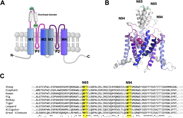
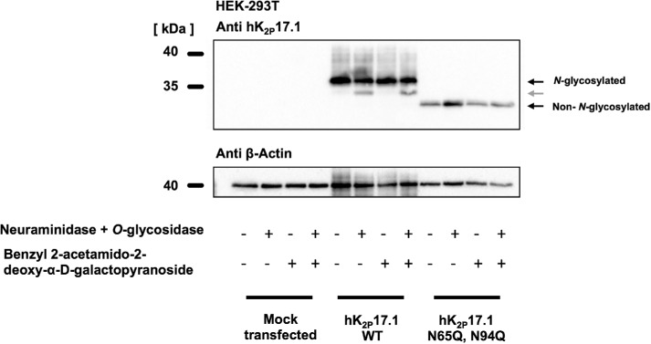
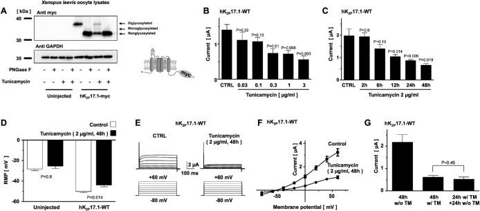
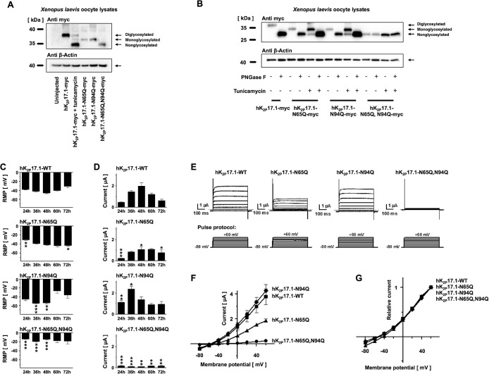
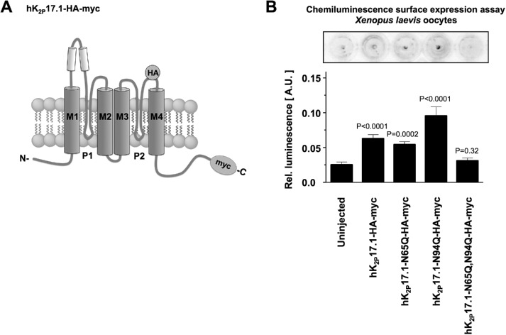
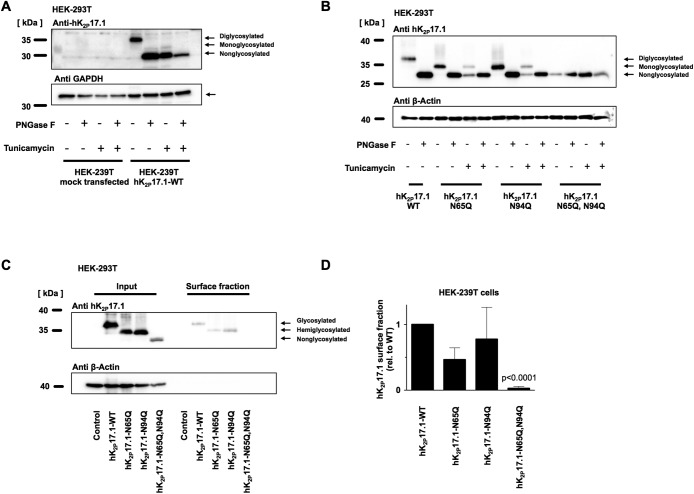
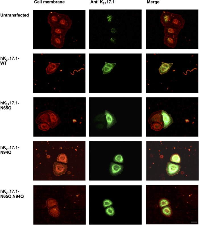
Two pore-domain potassium (K) channels mediate potassium background currents that stabilize the resting membrane potential and facilitate action potential repolarization. In the human heart, hK17.1 channels are predominantly expressed in the atria and Purkinje cells. Reduced atrial hK17.1 protein levels were described in patients with atrial fibrillation or heart failure. Genetic alterations in hK17.1 were associated with cardiac conduction disorders. Little is known about posttranslational modifications of hK17.1. Here, we characterized glycosylation of hK17.1 and investigated how glycosylation alters its surface expression and activity. Wild-type hK17.1 channels and channels lacking specific glycosylation sites were expressed in oocytes, HEK-293T cells, and HeLa cells. N-glycosylation was disrupted using N-glycosidase F and tunicamycin. hK17.1 expression and activity were assessed using immunoblot analysis and a two-electrode voltage clamp technique. Channel subunits of hK17.1 harbor two functional N-glycosylation sites at positions N65 and N94. In hemi-glycosylated hK17.1 channels, functionality and membrane trafficking remain preserved. Disruption of both N-glycosylation sites results in loss of hK17.1 currents, presumably caused by impaired surface expression. This study confirms diglycosylation of hK17.1 channel subunits and its pivotal role in cell-surface targeting. Our findings underline the functional relevance of N-glycosylation in biogenesis and membrane trafficking of ion channels.
双孔钾(K)通道介导钾背景电流,稳定静息膜电位并促进动作电位复极化。在人心肌中,hK17.1 通道主要在心房和浦肯野细胞中表达。在心房颤动或心力衰竭患者中,描述了心房 hK17.1 蛋白水平降低。hK17.1 的遗传改变与心脏传导障碍有关。关于 hK17.1 的翻译后修饰知之甚少。在这里,我们描述了 hK17.1 的糖基化,并研究了糖基化如何改变其表面表达和活性。野生型 hK17.1 通道和缺乏特定糖基化位点的通道在卵母细胞、HEK-293T 细胞和 HeLa 细胞中表达。使用 N-糖苷酶 F 和衣霉素破坏 N-糖基化。使用免疫印迹分析和双电极电压钳技术评估 hK17.1 的表达和活性。hK17.1 的通道亚基在位置 N65 和 N94 具有两个功能性 N-糖基化位点。在半糖基化的 hK17.1 通道中,功能和膜运输仍然保持。破坏两个 N-糖基化位点会导致 hK17.1 电流丧失,可能是由于表面表达受损所致。本研究证实了 hK17.1 通道亚基的双糖基化及其在细胞表面靶向中的关键作用。我们的发现强调了 N-糖基化在离子通道生物发生和膜运输中的功能相关性。