Department of Experimental Pharmacology, Mossakowski Medical Research Centre, Polish Academy of Sciences, Warsaw, Poland.
Small Animal Magnetic Resonance Imaging Laboratory, Mossakowski Medical Research Centre, Polish Academy of Sciences, Warsaw, Poland.
PLoS One. 2019 Apr 11;14(4):e0215348. doi: 10.1371/journal.pone.0215348. eCollection 2019.
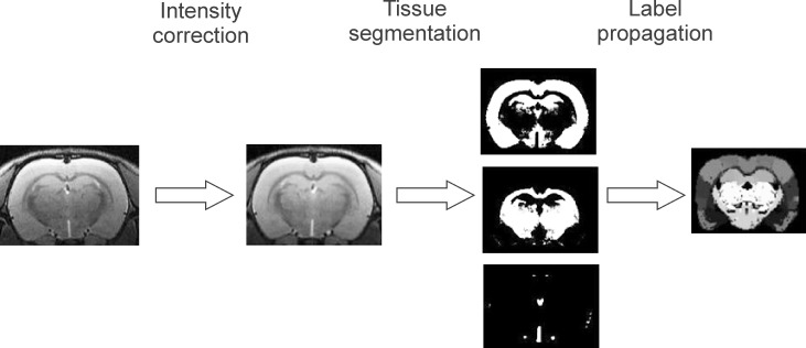
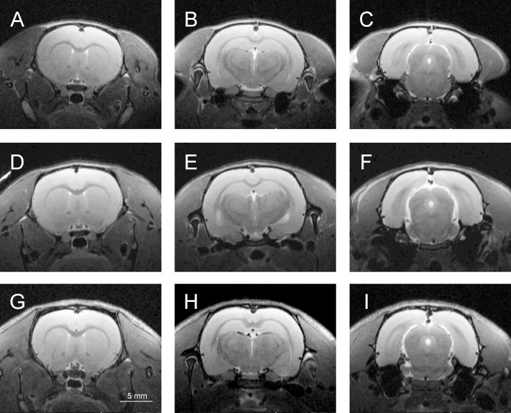
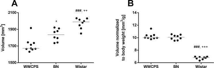
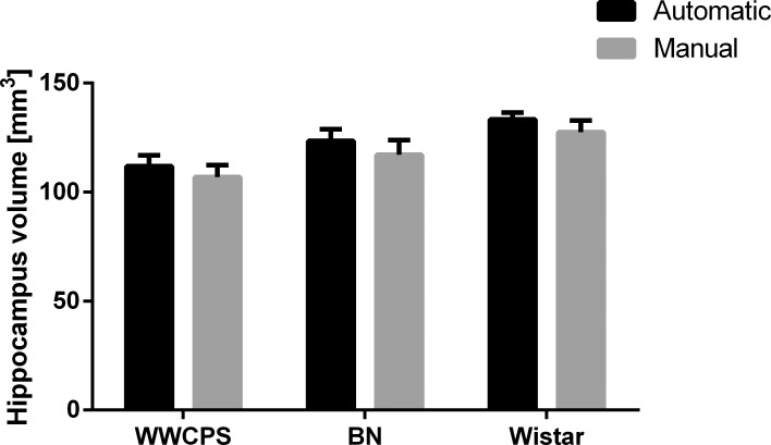
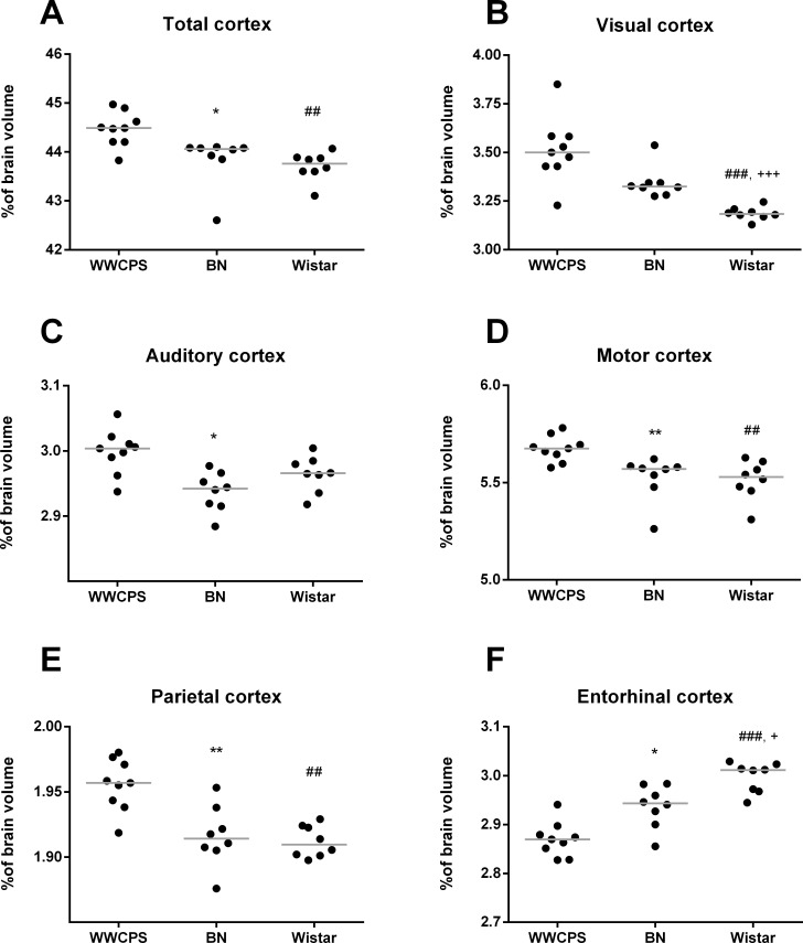
Selective breeding of laboratory rats resulted in changes of their behavior. Concomitantly, the albino strains developed vision related pathologies. These alterations certainly occurred on the background of modifications in brain morphology. The aim of the study was to assess and compare volumes of major structures in brains of wild-captive, laboratory albino and laboratory pigmented rats. High resolution T2-weighted images of brains of adult male Warsaw Wild Captive Pisula-Stryjek rats (WWCPS, a model of wild type), laboratory pigmented (Brown Norway strain, BN) and albino rats (Wistar strain, WI) were obtained with a 7T small animal-dedicated magnetic resonance tomograph. Volume quantification of whole brains and 50 brain structures within each brain were performed with the digital Schwarz rat brain atlas and a custom-made MATLAB/SPM8 scripts. Brain volumes were scaled to body mass, whereas volumes of brain structures were normalized to individual brain volumes. Normalized brain volume was similar in WWCPS and BN, but lower in WI. Normalized neocortex volume was smaller in both laboratory strains than in WWCPS and the visual cortex was smaller in albino WI rats than in WWCPS and BN. Relative volumes of phylogenetically older structures, such as hippocampus, amygdala, nucleus accumbens and olfactory nuclei, also displayed certain strain-related differences. The present data shows that selective breeding of laboratory rats markedly affected brain morphology, the neocortex being most significantly altered. In particular, albino rats display reduced volume of the visual cortex, possibly related to retinal degeneration and the development of blindness.
实验室大鼠的选择性繁殖导致其行为发生变化。同时,白化品系发展出与视觉相关的病变。这些改变肯定是在大脑形态改变的背景下发生的。本研究的目的是评估和比较野生-圈养、实验室白化和实验室色素大鼠大脑主要结构的体积。使用 7T 小动物专用磁共振断层扫描仪获得成年雄性华沙野生圈养 Pisula-Stryjek 大鼠(WWCPS,野生型模型)、实验室色素(棕色挪威品系,BN)和白化大鼠(Wistar 品系,WI)的大脑高分辨率 T2 加权图像。使用数字 Schwarz 大鼠脑图谱和定制的 MATLAB/SPM8 脚本对整个大脑和每个大脑中的 50 个脑结构进行体积量化。脑体积按体重进行缩放,而脑结构体积按个体脑体积进行归一化。WWCPS 和 BN 的标准化脑体积相似,但 WI 的标准化脑体积较低。两个实验室品系的新皮质体积均小于 WWCPS,白化 WI 大鼠的视觉皮质体积小于 WWCPS 和 BN。在进化上较老的结构,如海马体、杏仁核、伏隔核和嗅核的相对体积也显示出某些与品系相关的差异。目前的数据表明,实验室大鼠的选择性繁殖显著影响了大脑形态,新皮质的变化最为显著。特别是白化大鼠的视觉皮层体积减小,可能与视网膜退化和失明的发展有关。