Fonseka Pamali, Liem Michael, Ozcitti Cemil, Adda Christopher G, Ang Ching-Seng, Mathivanan Suresh
Department of Biochemistry and Genetics, La Trobe Institute for Molecular Science, La Trobe University, Melbourne, Australia.
The Bio21 Molecular Science and Biotechnology Institute, University of Melbourne, Parkville, Australia.
J Extracell Vesicles. 2019 Apr 11;8(1):1597614. doi: 10.1080/20013078.2019.1597614. eCollection 2019.
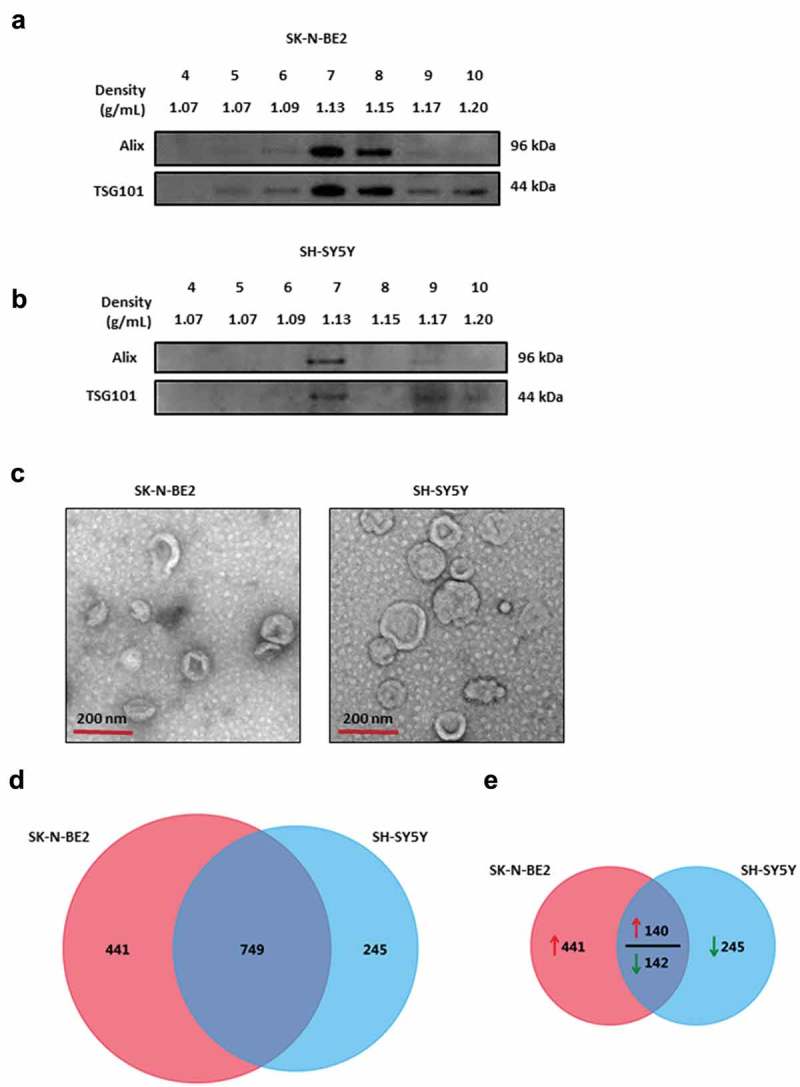
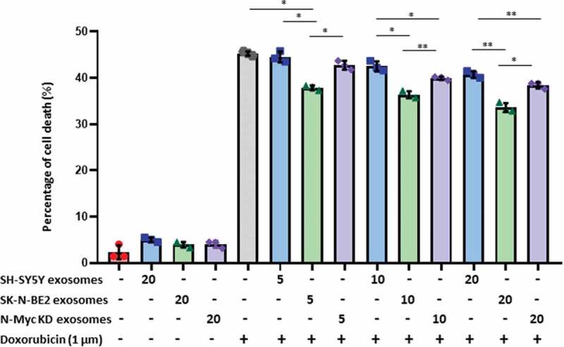
Neuroblastoma accounts for 15% of childhood cancer mortality. Amplification of the oncogene N-Myc is a well-established poor prognostic marker for neuroblastoma. Whilst N-Myc amplification status strongly correlates with higher tumour aggression and resistance to treatment, the role of N-Myc in the aggressiveness of the disease is poorly understood. Exosomes are released by many cell types including cancer cells and are implicated as key mediators in cell-cell communication via the transfer of molecular cargo. Hence, characterising the exosomal protein components from N-Myc amplified and non-amplified neuroblastoma cells will improve our understanding on their role in the progression of neuroblastoma. In this study, a comparative proteomic analysis of exosomes isolated from cells with varying N-Myc amplification status was performed. Label-free quantitative proteomic profiling revealed 968 proteins that are differentially abundant in exosomes released by the neuroblastoma cells. Gene ontology-based analysis highlighted the enrichment of proteins involved in cell communication and signal transduction in N-Myc amplified exosomes. Treatment of SH-SY5Y cells with N-Myc amplified SK-N-BE2 cell-derived exosomes increased the migratory potential, colony forming abilities and conferred resistance to doxorubicin induced apoptosis. Incubation of exosomes from N-Myc knocked down SK-N-BE2 cells abolished the transfer of resistance to doxorubicin induced apoptosis. These findings suggest that exosomes could play a pivotal role in N-Myc-driven aggressive neuroblastoma and transfer of chemoresistance between cells. : RNA = ribonucleic acid; DNA = deoxyribonucleic acid; FCS = foetal calf serum; NTA = nanoparticle tracking analysis; LC-MS = liquid chromatography-mass spectrometry; KD = knockdown; LTQ = linear trap quadropole; TEM = transmission electron microscopy.
神经母细胞瘤占儿童癌症死亡率的15%。癌基因N-Myc的扩增是神经母细胞瘤公认的不良预后标志物。虽然N-Myc扩增状态与更高的肿瘤侵袭性和治疗耐药性密切相关,但N-Myc在该疾病侵袭性中的作用仍知之甚少。外泌体由包括癌细胞在内的多种细胞类型释放,并被认为是通过分子货物转移进行细胞间通讯的关键介质。因此,表征N-Myc扩增和未扩增的神经母细胞瘤细胞的外泌体蛋白质成分将增进我们对它们在神经母细胞瘤进展中作用的理解。在本研究中,对从具有不同N-Myc扩增状态的细胞中分离出的外泌体进行了比较蛋白质组学分析。无标记定量蛋白质组学分析揭示了968种在神经母细胞瘤细胞释放的外泌体中丰度存在差异的蛋白质。基于基因本体的分析突出了N-Myc扩增外泌体中参与细胞通讯和信号转导的蛋白质的富集。用N-Myc扩增的SK-N-BE2细胞衍生的外泌体处理SH-SY5Y细胞可增加其迁移潜力、集落形成能力,并赋予对阿霉素诱导的凋亡的抗性。用N-Myc敲低的SK-N-BE2细胞的外泌体孵育可消除对阿霉素诱导的凋亡抗性的转移。这些发现表明,外泌体可能在N-Myc驱动的侵袭性神经母细胞瘤以及细胞间化疗耐药性转移中起关键作用。:RNA = 核糖核酸;DNA = 脱氧核糖核酸;FCS = 胎牛血清;NTA = 纳米颗粒跟踪分析;LC-MS = 液相色谱-质谱联用;KD = 敲低;LTQ = 线性离子阱;TEM = 透射电子显微镜。