• 文献检索
  • 文档翻译
  • 深度研究
  • 学术资讯
  • Suppr Zotero 插件Zotero 插件
  • 邀请有礼
  • 套餐&价格
  • 历史记录
应用&插件
Suppr Zotero 插件Zotero 插件浏览器插件Mac 客户端Windows 客户端微信小程序
定价
高级版会员购买积分包购买API积分包
服务
文献检索文档翻译深度研究API 文档MCP 服务
关于我们
关于 Suppr公司介绍联系我们用户协议隐私条款
关注我们

Suppr 超能文献

核心技术专利:CN118964589B侵权必究
粤ICP备2023148730 号-1Suppr @ 2026

文献检索

告别复杂PubMed语法,用中文像聊天一样搜索,搜遍4000万医学文献。AI智能推荐,让科研检索更轻松。

立即免费搜索

文件翻译

保留排版,准确专业,支持PDF/Word/PPT等文件格式,支持 12+语言互译。

免费翻译文档

深度研究

AI帮你快速写综述,25分钟生成高质量综述,智能提取关键信息,辅助科研写作。

立即免费体验

超声如何诊治胰腺疾病?超声表现与扫查手法。

How Does Ultrasound Manage Pancreatic Diseases? Ultrasound Findings and Scanning Maneuvers.

机构信息

Department of Gastroenterology, Iida Municipal Hospital, Iida, Japan.

出版信息

Gut Liver. 2020 Jan 15;14(1):37-46. doi: 10.5009/gnl18567.

DOI:10.5009/gnl18567
PMID:31009959
原文链接:https://pmc.ncbi.nlm.nih.gov/articles/PMC6974328/
Abstract

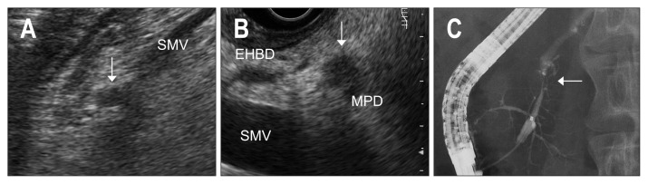
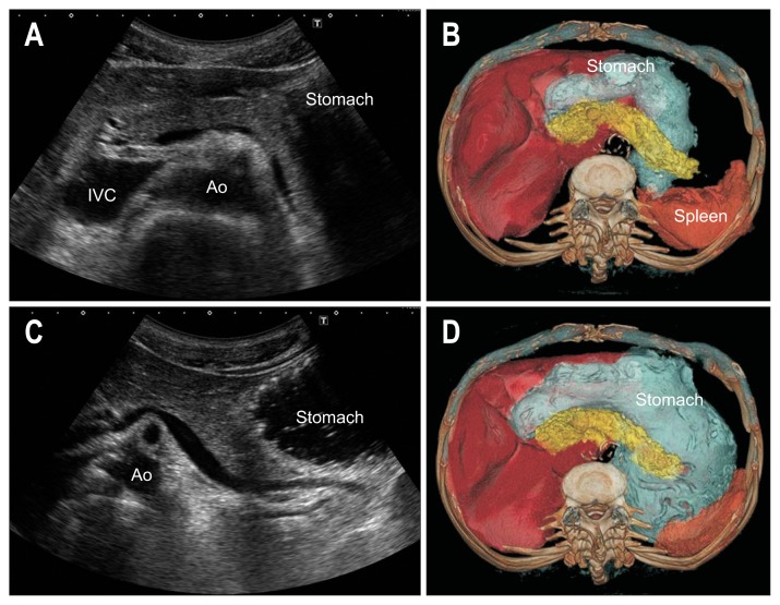
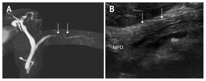
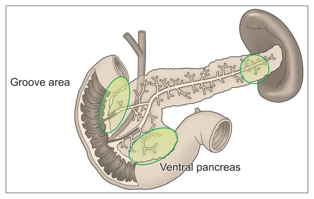
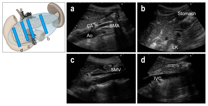
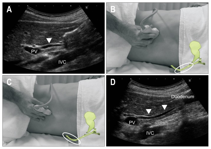
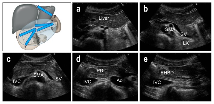
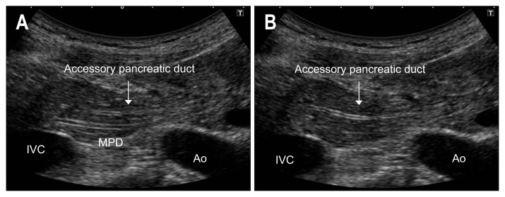
As ultrasound (US) is simple and less invasive than other imaging modalities, this technique is widely used for mass screening. However, visualizing the entire pancreas due to complicated anatomy, obesity and overlying gas can be difficult. US plays a key role in the diagnosis of pancreatic carcinoma (PC), of which tumors smaller than 10 mm (TS1a) and pancreatic carcinoma are expected to have good prognosis. To detect these forms of PC, main pancreatic duct (MPD) dilatation (3 mm or more) and pancreatic cysts (5 mm or larger) are US findings of high-risk individuals (HRIs), and these subjects should be observed periodically. Scanning maneuvers are also important for both screening for PC and follow-up of HRIs. As lesions in the groove area and ventral pancreas do not affect the MPD or extrahepatic bile duct, we should pay attention to these areas. Visualization of the tail is also challenging due to gas and stool in the alimentary tract. As the position of the pancreas changes depending on the body posture, and several different body positions should be employed, such as the right lateral decubitus, sitting, and upright positions, rather than only applying strong compression with the transducer. In cases with poor visualization, the liquid-filled stomach method is highly recommended.

摘要

由于超声(US)比其他成像方式简单且侵入性更小,因此该技术被广泛用于大规模筛查。然而,由于胰腺的复杂解剖结构、肥胖和上方气体的影响,很难对整个胰腺进行可视化。US 在胰腺癌(PC)的诊断中起着关键作用,其中直径小于 10mm(TS1a)和胰腺神经内分泌肿瘤的肿瘤预计具有良好的预后。为了检测这些形式的 PC,主胰管(MPD)扩张(3mm 或更大)和胰腺囊肿(5mm 或更大)是高危人群(HRIs)的 US 发现,这些患者应定期观察。扫描手法对于 PC 的筛查和 HRIs 的随访也很重要。由于沟区和胰头区域的病变不会影响胰管或肝外胆管,因此我们应该注意这些区域。由于消化道中的气体和粪便,尾部的可视化也具有挑战性。由于胰腺的位置随体位变化而变化,因此应采用几种不同的体位,如右侧卧位、坐位和立位,而不是仅用探头进行强力压迫。在可视化效果不佳的情况下,强烈推荐使用充满液体的胃法。

https://cdn.ncbi.nlm.nih.gov/pmc/blobs/7b55/6974328/aff7d9933bb7/gnl-14-037f12.jpg
https://cdn.ncbi.nlm.nih.gov/pmc/blobs/7b55/6974328/dde3740c21a5/gnl-14-037f1.jpg
https://cdn.ncbi.nlm.nih.gov/pmc/blobs/7b55/6974328/79e12d54b1f6/gnl-14-037f2.jpg
https://cdn.ncbi.nlm.nih.gov/pmc/blobs/7b55/6974328/70c13795ea25/gnl-14-037f3.jpg
https://cdn.ncbi.nlm.nih.gov/pmc/blobs/7b55/6974328/0a5379b49965/gnl-14-037f4.jpg
https://cdn.ncbi.nlm.nih.gov/pmc/blobs/7b55/6974328/3b0584f21d47/gnl-14-037f5.jpg
https://cdn.ncbi.nlm.nih.gov/pmc/blobs/7b55/6974328/49547ed0835c/gnl-14-037f6.jpg
https://cdn.ncbi.nlm.nih.gov/pmc/blobs/7b55/6974328/b30f873e79ee/gnl-14-037f7.jpg
https://cdn.ncbi.nlm.nih.gov/pmc/blobs/7b55/6974328/fdad96ee8cf8/gnl-14-037f8.jpg
https://cdn.ncbi.nlm.nih.gov/pmc/blobs/7b55/6974328/c21d858c9f9a/gnl-14-037f9.jpg
https://cdn.ncbi.nlm.nih.gov/pmc/blobs/7b55/6974328/18c7bb9ae15c/gnl-14-037f10.jpg
https://cdn.ncbi.nlm.nih.gov/pmc/blobs/7b55/6974328/bc3ef4a12623/gnl-14-037f11.jpg
https://cdn.ncbi.nlm.nih.gov/pmc/blobs/7b55/6974328/aff7d9933bb7/gnl-14-037f12.jpg

相似文献

1
How Does Ultrasound Manage Pancreatic Diseases? Ultrasound Findings and Scanning Maneuvers.超声如何诊治胰腺疾病?超声表现与扫查手法。
Gut Liver. 2020 Jan 15;14(1):37-46. doi: 10.5009/gnl18567.
2
Endoscopic Ultrasound-Based Pancreatic Cancer Screening of High-Risk Individuals: A Prospective Observational Trial.基于内镜超声的高危个体胰腺癌筛查:一项前瞻性观察性试验。
Pancreas. 2018 May/Jun;47(5):586-591. doi: 10.1097/MPA.0000000000001038.
3
Cost-effectiveness of Abdominal Ultrasound Versus Magnetic Resonance Imaging for Pancreatic Cancer Screening in Familial High-Risk Individuals in Japan.日本家族性高危人群胰腺癌筛查中腹部超声与磁共振成像的成本效益比较。
Pancreas. 2020 Sep;49(8):1052-1056. doi: 10.1097/MPA.0000000000001614.
4
Morphologic changes in the pancreas detected by screening ultrasonography in a mass survey, with special reference to main duct dilatation, cyst formation, and calcification.在大规模调查中通过筛查超声检查检测到的胰腺形态学变化,特别提及主胰管扩张、囊肿形成和钙化。
Pancreas. 1994 Jul;9(4):508-12. doi: 10.1097/00006676-199407000-00014.
5
Prospective Follow-up Study of the Recurrence of Pancreatic Cancer Diagnosed at an Early Stage: The Value of Endoscopic Ultrasonography for Early Diagnosis of Recurrence in the Remnant Pancreas.早期诊断的胰腺癌复发的前瞻性随访研究:内镜超声对残胰复发早期诊断的价值。
Pancreas. 2018 Apr;47(4):482-488. doi: 10.1097/MPA.0000000000001021.
6
Feasibility of a complete pancreatobiliary linear endoscopic ultrasound examination from the stomach.从胃进行全胰胆线性内镜超声检查的可行性。
Endoscopy. 2018 Jan;50(1):22-32. doi: 10.1055/s-0043-118592. Epub 2017 Sep 21.
7
Computed tomography of the pancreas: three second scanning versus 18 second scanning.胰腺的计算机断层扫描:3秒扫描与18秒扫描
J Comput Assist Tomogr. 1982 Apr;6(2):259-67. doi: 10.1097/00004728-198204000-00006.
8
A multicentre comparative prospective blinded analysis of EUS and MRI for screening of pancreatic cancer in high-risk individuals.EUS 和 MRI 用于高危人群胰腺癌筛查的多中心前瞻性盲法比较分析。
Gut. 2016 Sep;65(9):1505-13. doi: 10.1136/gutjnl-2014-308008. Epub 2015 May 18.
9
Intraoperative ultrasonography by high-resolution annular array transducer for intraductal papillary mucinous tumors of the pancreas.使用高分辨率环形阵列换能器对胰腺导管内乳头状黏液性肿瘤进行术中超声检查。
Surgery. 2001 Jan;129(1):55-65. doi: 10.1067/msy.2001.109118.
10
Ultrasonography of the pancreas. 1. Conventional imaging.胰腺超声检查。1. 传统成像。
Abdom Imaging. 2007 Mar-Apr;32(2):136-49. doi: 10.1007/s00261-006-9079-y.

引用本文的文献

1
Advanced ultrasound diagnosis of extrahepatic bile duct lesions.肝外胆管病变的超声高级诊断
J Med Ultrason (2001). 2025 Jan;52(1):69-83. doi: 10.1007/s10396-024-01491-3. Epub 2024 Oct 21.
2
Pancreatic duct imaging during aging.衰老过程中的胰管成像。
Endosc Ultrasound. 2023 Mar-Apr;12(2):200-212. doi: 10.4103/EUS-D-22-00119.
3
Two Cases of Perihepatitis With the Liver Capsule Irritation Sign: A New Physical Examination Technique.两例伴有肝包膜刺激征的肝周炎:一种新的体格检查技术

本文引用的文献

1
Multicenter study of early pancreatic cancer in Japan.日本多中心早期胰腺癌研究。
Pancreatology. 2018 Jan;18(1):61-67. doi: 10.1016/j.pan.2017.11.007. Epub 2017 Nov 20.
2
Evaluating the ability to detect pancreatic lesions using a special ultrasonography examination focusing on the pancreas.通过聚焦胰腺的特殊超声检查评估检测胰腺病变的能力。
Eur J Radiol. 2017 Jun;91:10-14. doi: 10.1016/j.ejrad.2017.03.010. Epub 2017 Mar 22.
3
Clinical Practice Guidelines for Pancreatic Cancer 2016 From the Japan Pancreas Society: A Synopsis.
Cureus. 2023 Jan 29;15(1):e34327. doi: 10.7759/cureus.34327. eCollection 2023 Jan.
4
Carcinoma in situ of the pancreas with fibrosis area around the carcinoma: A case report.胰腺原位癌伴癌周纤维化区域:1例报告
Medicine (Baltimore). 2020 Oct 16;99(42):e22645. doi: 10.1097/MD.0000000000022645.
《2016年日本胰腺学会胰腺癌临床实践指南:概要》
Pancreas. 2017 May/Jun;46(5):595-604. doi: 10.1097/MPA.0000000000000816.
4
Ultrasonographic diagnostic criteria for pancreatic cancer.胰腺癌的超声诊断标准。
J Med Ultrason (2001). 2013 Oct;40(4):497-504. doi: 10.1007/s10396-013-0483-y.
5
Pancreatic cysts in general population on ultrasonography: Prevalence and development of risk score.超声检查一般人群中的胰腺囊肿:患病率及风险评分的发展情况
J Gastroenterol. 2016 Dec;51(12):1133-1140. doi: 10.1007/s00535-016-1196-y. Epub 2016 Mar 17.
6
Pancreatic cancer.胰腺癌。
Lancet. 2016 Jul 2;388(10039):73-85. doi: 10.1016/S0140-6736(16)00141-0. Epub 2016 Jan 30.
7
Diagnostic strategies for early pancreatic cancer.早期胰腺癌的诊断策略
J Gastroenterol. 2015 Feb;50(2):147-54. doi: 10.1007/s00535-014-1026-z. Epub 2014 Dec 14.
8
Preliminary study on evaluation of the pancreatic tail observable limit of transabdominal ultrasonography using a position sensor and CT-fusion image.利用位置传感器和CT融合图像评估经腹超声对胰尾可观察限度的初步研究
Eur J Radiol. 2014 Aug;83(8):1324-31. doi: 10.1016/j.ejrad.2014.05.009. Epub 2014 May 19.
9
Cancer statistics, 2014.癌症统计数据,2014 年。
CA Cancer J Clin. 2014 Jan-Feb;64(1):9-29. doi: 10.3322/caac.21208. Epub 2014 Jan 7.
10
The pancreatic head is larger than often assumed.胰头比通常认为的要大。
Z Gastroenterol. 2013 Apr;51(4):390-4. doi: 10.1055/s-0033-1335175. Epub 2013 Apr 12.