Division of Pathology and Cell Therapy, Chiba Cancer Center Research Institute, Japan.
Laboratory of Molecular Cell Biology, Graduate School of Pharmaceutical Sciences, Chiba University, Japan.
Mol Oncol. 2019 Jun;13(6):1419-1432. doi: 10.1002/1878-0261.12496. Epub 2019 May 15.
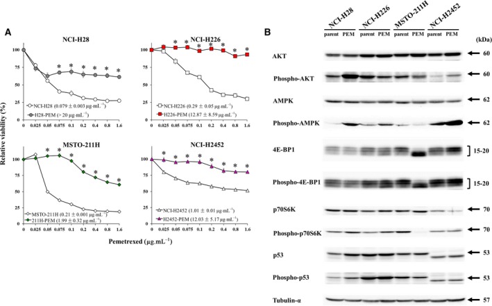
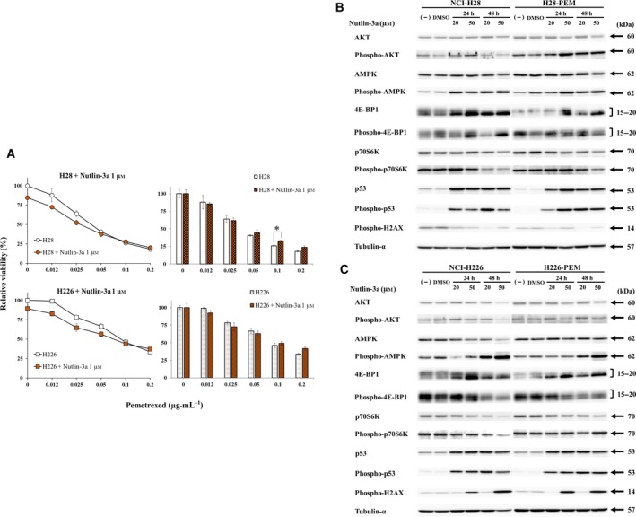
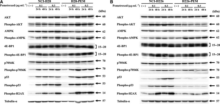
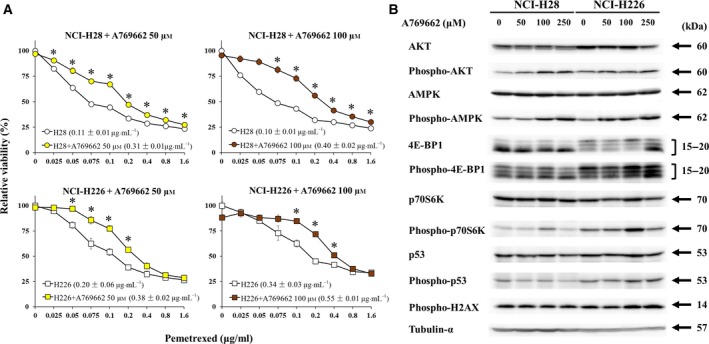
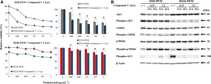
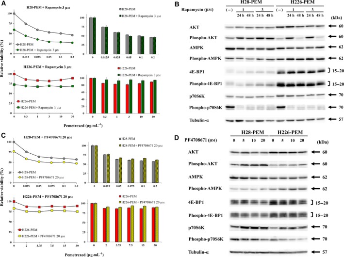
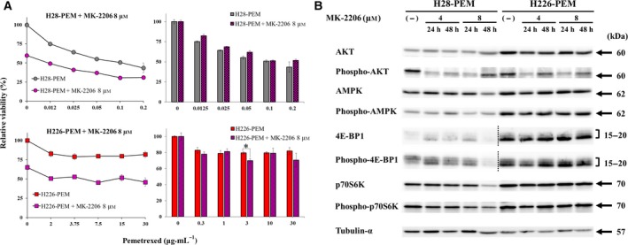
Pemetrexed (PEM) inhibits DNA and RNA synthesis and is currently one of the first-line agents for mesothelioma. PEM suppresses the activities of several enzymes involved in purine and pyrimidine synthesis, and elevated activity of these enzymes in tumors is often linked with resistance to PEM. The agent also stimulates AMP-activated protein kinase (AMPK) and consequently influences the mammalian target of rapamycin complex 1 (mTORC1) pathways. Nevertheless, it remains unclear whether PEM resistance is linked to the AMPK or mTORC1 pathways. Here, we established two independent PEM-resistant mesothelioma cell lines in which expression of the PEM-target enzymes was not elevated, and found that levels of phosphorylated AMPK and p70S6K and, to a lesser extent, levels of phosphorylated AKT and p53, were increased in these cells as compared with the respective parent cells. PEM stimulation also augmented phosphorylation of AMPK, p70S6K, AKT and p53 in most cases. An AMPK activator increased phosphorylation and PEM resistance in parental cells, and the inhibitor decreased the resistance of PEM-resistant cells. In contrast, inhibitors for p70S6K and AKT did not influence PEM resistance; furthermore, increased levels of endogenous p53 did not affect PEM sensitivity. These data collectively indicate that constitutive activation of AMPK is associated with PEM resistance, and that this is unconnected with elevated DNA and RNA synthesis.
培美曲塞(PEM)抑制 DNA 和 RNA 的合成,目前是间皮瘤的一线治疗药物之一。PEM 抑制几种参与嘌呤和嘧啶合成的酶的活性,而这些酶在肿瘤中的高活性通常与对 PEM 的耐药性有关。该药物还刺激 AMP 激活的蛋白激酶(AMPK),进而影响雷帕霉素靶蛋白复合物 1(mTORC1)途径。然而,目前尚不清楚 PEM 耐药性是否与 AMPK 或 mTORC1 途径有关。在这里,我们建立了两个独立的 PEM 耐药性间皮瘤细胞系,其中 PEM 靶酶的表达没有升高,我们发现这些细胞中磷酸化 AMPK 和 p70S6K 的水平升高,在一定程度上,磷酸化 AKT 和 p53 的水平也升高,与相应的亲本细胞相比。PEM 刺激也增加了 AMPK、p70S6K、AKT 和 p53 的磷酸化。在大多数情况下,AMPK 激活剂增加了亲本细胞的磷酸化和 PEM 耐药性,而抑制剂降低了 PEM 耐药细胞的耐药性。相比之下,p70S6K 和 AKT 的抑制剂并不影响 PEM 耐药性;此外,内源性 p53 水平的增加并不影响 PEM 的敏感性。这些数据表明,AMPK 的组成性激活与 PEM 耐药性有关,与 DNA 和 RNA 合成的升高无关。