Mousazadeh Sepideh, Ghaheri Azadeh, Shahhoseini Maryam, Aflatoonian Reza, Afsharian Parvaneh
Department of Genetics, School of Natural Sciences, University of Tabriz, Tabriz, Iran.
Department of Genetics, Reproductive Biomedicine Research Center, Royan Institute for Reproductive Biomedicine, ACECR, Tehran, Iran.
Int J Fertil Steril. 2019 Jul;13(2):127-134. doi: 10.22074/ijfs.2019.5604. Epub 2019 Apr 27.
Gelatinases degrade extracellular matrix (ECM) components to allow for physiological remodeling and contribute to pathological tissue destruction in endometriosis. It is known that the function of gelatinases is resistant to suppression by progesterone in endometriosis. The ability of progesterone to impact gene expression depends on the ratio. An imbalanced ratio in endometriotic tissue may be the result of the differential expression of and , which could be important in the etiology and pathogenesis of the disease. Hence, we decided to study the association of ratio and gelatinases expression in endometriosis.
In this prospective case-control study, we enrolled 40 women, 20 in the case group who were diagnosed with stage III/IV endometriosis and 20 normal subjects without endometriosis (controls) who referred to Royan Institute, Tehran, Iran during 2013-2014. We obtained 60 tissue samples [ectopic (n=20), eutopic (n=20), and normal endometrium (n=20)]. RNA was extracted from the tissue samples in order to analyze , , , and mRNA levels through real-time polymerase chain reaction (PCR).
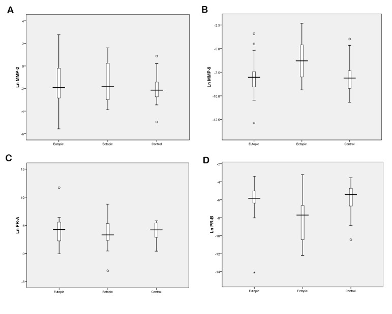
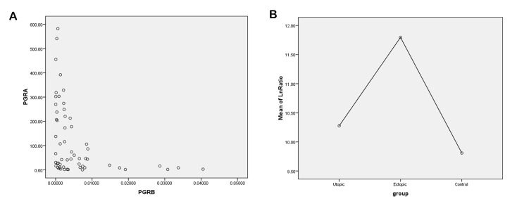
There was significantly lower expression of the isoform in ectopic tissues compared to the control (P=0.002) and eutopic endometrium (P=0.006) tissues. PR-A expression was higher, but not significantly so, in the same ectopic and eutopic endometrium tissues compared to the control tissues (P=0.643). There was significant overexpression of in ectopic samples compared to control (P=0.014) and eutopic endometrium (P=0.012) samples. The ratio was not significantly higher in either eutopic or ectopic samples compared to the control samples (P=0.305).
Our findings support an altered expression in endometriosis, which may be associated with overexpression. This finding can be important for disease pathogenesis.
明胶酶可降解细胞外基质(ECM)成分,以实现生理重塑,并参与子宫内膜异位症中的病理性组织破坏。已知在子宫内膜异位症中,明胶酶的功能对孕激素的抑制具有抗性。孕激素影响基因表达的能力取决于[此处原文缺失相关比值内容]的比例。子宫内膜异位组织中[此处原文缺失相关比值内容]比例失衡可能是[此处原文缺失相关基因内容]和[此处原文缺失相关基因内容]差异表达的结果,这可能在该疾病的病因和发病机制中具有重要意义。因此,我们决定研究子宫内膜异位症中[此处原文缺失相关比值内容]比例与明胶酶表达之间的关联。
在这项前瞻性病例对照研究中,我们纳入了40名女性,其中病例组20名,被诊断为III/IV期子宫内膜异位症;20名正常受试者(对照组),未患子宫内膜异位症。这些受试者于2013 - 2014年期间前往伊朗德黑兰的罗扬研究所就诊。我们获取了60份组织样本[异位内膜(n = 20)、在位内膜(n = 20)和正常子宫内膜(n = 20)]。从组织样本中提取RNA,以便通过实时聚合酶链反应(PCR)分析[此处原文缺失相关基因内容]、[此处原文缺失相关基因内容]、[此处原文缺失相关基因内容]和[此处原文缺失相关基因内容]的mRNA水平。
与对照组(P = 0.002)和在位内膜组织(P = 0.006)相比,异位组织中[此处原文缺失相关基因内容]同工型的表达显著降低。与对照组织相比,相同的异位和在位内膜组织中PR - A表达较高,但差异不显著(P = 0.643)。与对照组(P = 0.014)和在位内膜样本(P = 0.012)相比,异位样本中[此处原文缺失相关基因内容]有显著过表达。与对照样本相比,在位或异位样本中的[此处原文缺失相关比值内容]比例均未显著升高(P = 0.305)。
我们的研究结果支持子宫内膜异位症中[此处原文缺失相关基因内容]表达改变,这可能与[此处原文缺失相关基因内容]过表达有关。这一发现可能对疾病发病机制具有重要意义。