Department of Neurology (S.K., W.B., M.E.) and Department of Neuropathology (C.P., N.F., H.R., R.M., V.M., H.-H.G., W.S.), Charité-Universitätsmedizin, Berlin, Germany; Department of Internal Medicine and Clinical Immunology (Y.A., O.B.), Assistance Public-Hôpitaux de Paris, Sorbonne-Université, INSERM, UMR974, Pitié-Salpêtrière University Hospital; Unité de Pathologie Neuromusculaire (S.L.-L.), Centre de Référence Paris-Est, Groupe Hospitalier Pitié-Salpêtrière; Service de Neurologie 2-Mazarin (M.T.), Hôpitaux Universitaires La Pitié Salpêtrière-Charles Foix, APHP; OncoNeuroTox Group (M.T.), Center for Patients with Neurological Complications of Oncologic Treatments, Hôpitaux Universitaires Pitié-Salpetrière-Charles Foix et Hôpital Percy; Inserm U 1127 (M.T.), CNRS UMR 7225, Institut du Cerveau et de la Moelle épinière, ICM, Université Pierre-et-Marie-Curie, Sorbonne Université, Paris, France; Leibniz ScienceCampus Chronic Inflammation (H.R., R.M., W.S.); Center for Stroke Research Berlin (M.E.), Charité-Universitätsmedizin, Berlin; German Centre for Cardiovascular Research (DZHK) (M.E.); and German Center for Neurodegenerative Diseases (DZNE) (M.E.).
Neurol Neuroimmunol Neuroinflamm. 2019 Apr 10;6(3):e558. doi: 10.1212/NXI.0000000000000558. eCollection 2019 May.
To investigate the relevance of dysfunctional T cells in immune-mediated myopathies. We analyzed T-cell exhaustion and senescence, in the context of programmed cell death protein 1 (PD1)-related immunity in skeletal muscle biopsies from patients with immune-mediated necrotizing myopathy (IMNM), sporadic inclusion body myositis (sIBM), and myositis induced by immune checkpoint inhibitors (irMyositis).
Skeletal muscle biopsies from 12 patients with IMNM, 7 patients with sIBM, and 8 patients with irMyositis were analyzed by immunostaining and immunofluorescence as well as by quantitative PCR. Eight biopsies from nondisease participants served as controls.
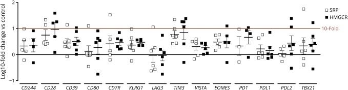
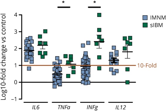
CD3CD8 T cells in biopsies from IMNM, sIBM, and irMyositis were largely PD1-positive, while CD68 macrophages were sparsely positive to the ligand of programmed cell death protein 1 (PD-L1). The sarcolemma of myofibers was PD-L2 and was colocalized with major histocompatibility complex (MHC) class I. CD68 macrophages were colocalized with PD-L2. Senescent T cells were strongly enriched in skeletal muscle of sIBM, revealing a distinct immunologic signature. Biopsies from patients with irMyositis showed mild signs of senescence and exhaustion.
Persistent exposure to antigens in IMNMs and sIBM may lead to T-cell exhaustion, a process controlled by the PD1 receptor and its cognate ligands PD-L1/PD-L2. To our knowledge, these data are the first evidence of presence of dysfunctional T cells and relevance of the PD1 pathway in IMNM, sIBM, and irMyositis. These findings may guide the way to a novel understanding of the immune pathogenesis of immune-mediated myopathies.
研究功能失调 T 细胞与免疫介导性肌病的相关性。我们分析了程序性细胞死亡蛋白 1(PD1)相关免疫中 T 细胞耗竭和衰老的情况,研究对象为免疫介导性坏死性肌病(IMNM)、散发性包涵体肌炎(sIBM)和免疫检查点抑制剂诱导的肌炎(irMyositis)患者的骨骼肌活检。
对 12 例 IMNM、7 例 sIBM 和 8 例 irMyositis 患者的骨骼肌活检进行免疫染色和免疫荧光分析以及定量 PCR 分析。8 例非疾病参与者的活检作为对照。
IMNM、sIBM 和 irMyositis 活检中的 CD3CD8 T 细胞大多为 PD1 阳性,而 CD68 巨噬细胞 PD1 配体(PD-L1)阳性稀疏。肌纤维的肌膜表达 PD-L2,与主要组织相容性复合体(MHC)I 类共定位。CD68 巨噬细胞与 PD-L2 共定位。sIBM 骨骼肌中衰老 T 细胞明显富集,呈现出独特的免疫特征。irMyositis 患者的活检显示出轻微的衰老和耗竭迹象。
在 IMNMs 和 sIBM 中,持续暴露于抗原可能导致 T 细胞耗竭,这是一个由 PD1 受体及其配体 PD-L1/PD-L2 控制的过程。据我们所知,这些数据是首次证明在 IMNM、sIBM 和 irMyositis 中存在功能失调的 T 细胞和 PD1 通路相关性的证据。这些发现可能为免疫介导性肌病的免疫发病机制提供新的认识。