• 文献检索
  • 文档翻译
  • 深度研究
  • 学术资讯
  • Suppr Zotero 插件Zotero 插件
  • 邀请有礼
  • 套餐&价格
  • 历史记录
应用&插件
Suppr Zotero 插件Zotero 插件浏览器插件Mac 客户端Windows 客户端微信小程序
定价
高级版会员购买积分包购买API积分包
服务
文献检索文档翻译深度研究API 文档MCP 服务
关于我们
关于 Suppr公司介绍联系我们用户协议隐私条款
关注我们

Suppr 超能文献

核心技术专利:CN118964589B侵权必究
粤ICP备2023148730 号-1Suppr @ 2026

文献检索

告别复杂PubMed语法,用中文像聊天一样搜索,搜遍4000万医学文献。AI智能推荐,让科研检索更轻松。

立即免费搜索

文件翻译

保留排版,准确专业,支持PDF/Word/PPT等文件格式,支持 12+语言互译。

免费翻译文档

深度研究

AI帮你快速写综述,25分钟生成高质量综述,智能提取关键信息,辅助科研写作。

立即免费体验

鉴定视网膜钠钾-ATP 酶上与视蛋白结合的位点。

Identification of the retinoschisin-binding site on the retinal Na/K-ATPase.

机构信息

Institute of Human Genetics, University of Regensburg, Regensburg, Germany.

Institute of Biophysics and Physical Biochemistry, University of Regensburg, Regensburg, Germany.

出版信息

PLoS One. 2019 May 2;14(5):e0216320. doi: 10.1371/journal.pone.0216320. eCollection 2019.

DOI:10.1371/journal.pone.0216320
PMID:31048931
原文链接:https://pmc.ncbi.nlm.nih.gov/articles/PMC6497308/
Abstract

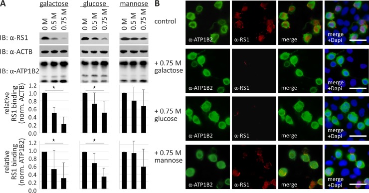
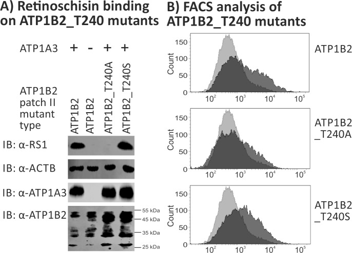
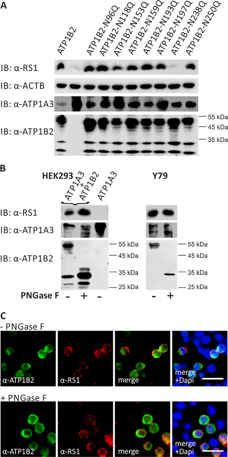
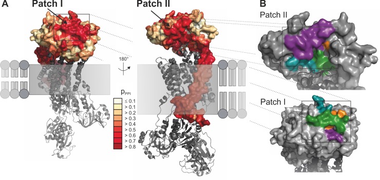
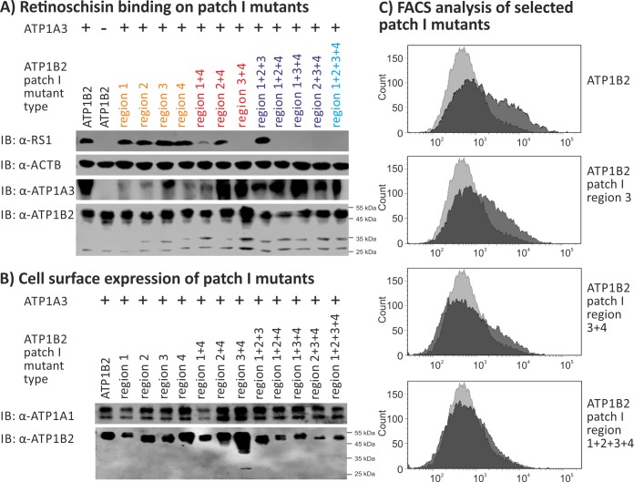
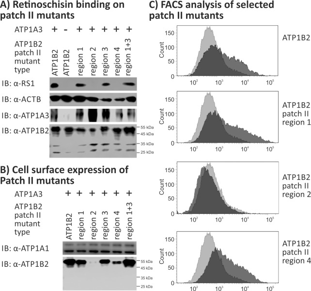
X-linked juvenile retinoschisis (XLRS) is a hereditary retinal dystrophy, caused by mutations in the RS1 gene which encodes the secreted protein retinoschisin. In recent years, several molecules have been proposed to interact with retinoschisin, including the retinal Na/K-ATPase, L-voltage gated Ca2+ channels, and specific sugars. We recently showed that the retinal Na/K-ATPase consisting of subunits ATP1A3 and ATP1B2 is essential for anchoring retinoschisin to plasma membranes and identified the glycosylated ATP1B2 subunit as the direct interaction partner for retinoschisin. We now aimed to precisely map the retinoschisin binding domain(s) in ATP1B2. In general, retinoschisin binding was not affected after selective elimination of individual glycosylation sites via site-directed mutagenesis as well as after full enzymatic deglycosylation of ATP1B2. Applying the interface prediction tool PresCont, two putative protein-protein interaction patches ("patch I" and "patch II") consisting each of four hydrophobic amino acid stretches on the ATP1B2 surface were identified. These were consecutively altered by site-directed mutagenesis. Functional assays with the ATP1B2 patch mutants identified patch II and, specifically, the associated amino acid at position 240 (harboring a threonine in ATP1B2) as crucial for retinoschisin binding to ATP1B2. These and previous results led us to suggest an induced-fit binding mechanism for the interaction between retinoschisin and the Na/K-ATPase, which is dependent on threonine 240 in ATP1B2 allowing the accommodation of hyperflexible retinoschisin spikes by the associated protein-protein interaction patch on ATP1B2.

摘要

X 连锁青少年性视网膜劈裂症(XLRS)是一种遗传性视网膜营养不良,由 RS1 基因的突变引起,该基因编码分泌蛋白视网膜蛋白聚糖。近年来,有几种分子被提出与视网膜蛋白聚糖相互作用,包括视网膜 Na/K-ATP 酶、L-电压门控 Ca2+通道和特定的糖。我们最近表明,由 ATP1A3 和 ATP1B2 亚基组成的视网膜 Na/K-ATP 酶对于将视网膜蛋白聚糖锚定在质膜上是必不可少的,并确定糖基化的 ATP1B2 亚基是与视网膜蛋白聚糖的直接相互作用伙伴。我们现在的目标是精确定位 ATP1B2 中与视网膜蛋白聚糖结合的(多个)结构域。一般来说,通过定点突变选择性消除单个糖基化位点后,以及在 ATP1B2 完全酶糖基化后,视网膜蛋白聚糖的结合没有受到影响。应用界面预测工具 PresCont,在 ATP1B2 表面上确定了两个由四个疏水性氨基酸片段组成的假定蛋白-蛋白相互作用斑块(“斑块 I”和“斑块 II”)。这些依次通过定点突变进行改变。对 ATP1B2 斑块突变体的功能测定确定了斑块 II,特别是与位置 240 相关的氨基酸(在 ATP1B2 中含有苏氨酸)对于视网膜蛋白聚糖与 ATP1B2 的结合至关重要。这些和以前的结果使我们提出了一种诱导契合结合机制,用于视网膜蛋白聚糖与 Na/K-ATP 酶之间的相互作用,该机制依赖于 ATP1B2 中的苏氨酸 240,允许相关的蛋白-蛋白相互作用斑块在 ATP1B2 上容纳超柔性的视网膜蛋白聚糖刺。

https://cdn.ncbi.nlm.nih.gov/pmc/blobs/b30e/6497308/37029d16882f/pone.0216320.g006.jpg
https://cdn.ncbi.nlm.nih.gov/pmc/blobs/b30e/6497308/945328350618/pone.0216320.g001.jpg
https://cdn.ncbi.nlm.nih.gov/pmc/blobs/b30e/6497308/02545f432e50/pone.0216320.g002.jpg
https://cdn.ncbi.nlm.nih.gov/pmc/blobs/b30e/6497308/ebad2a8617e4/pone.0216320.g003.jpg
https://cdn.ncbi.nlm.nih.gov/pmc/blobs/b30e/6497308/3d9768ee9d2a/pone.0216320.g004.jpg
https://cdn.ncbi.nlm.nih.gov/pmc/blobs/b30e/6497308/abe8fe20d909/pone.0216320.g005.jpg
https://cdn.ncbi.nlm.nih.gov/pmc/blobs/b30e/6497308/37029d16882f/pone.0216320.g006.jpg

相似文献

1
Identification of the retinoschisin-binding site on the retinal Na/K-ATPase.鉴定视网膜钠钾-ATP 酶上与视蛋白结合的位点。
PLoS One. 2019 May 2;14(5):e0216320. doi: 10.1371/journal.pone.0216320. eCollection 2019.
2
The Na/K-ATPase is obligatory for membrane anchorage of retinoschisin, the protein involved in the pathogenesis of X-linked juvenile retinoschisis.钠钾 ATP 酶是视锥视蛋白固定在细胞膜上所必需的,该蛋白与 X 连锁青年性视网膜劈裂症的发病机制有关。
Hum Mol Genet. 2011 Mar 15;20(6):1132-42. doi: 10.1093/hmg/ddq557. Epub 2010 Dec 31.
3
Retinoschisin is linked to retinal Na/K-ATPase signaling and localization.视网膜分裂蛋白与视网膜钠钾ATP酶信号传导及定位有关。
Mol Biol Cell. 2017 Aug 1;28(16):2178-2189. doi: 10.1091/mbc.E17-01-0064. Epub 2017 Jun 14.
4
Pathomechanism of mutated and secreted retinoschisin in X-linked juvenile retinoschisis.X 连锁青年性视网膜劈裂症中突变和分泌的视网膜色素上皮衍生因子的发病机制。
Exp Eye Res. 2018 Dec;177:23-34. doi: 10.1016/j.exer.2018.07.021. Epub 2018 Jul 21.
5
Retinoschisin and novel Na/K-ATPase interaction partners Kv2.1 and Kv8.2 define a growing protein complex at the inner segments of mammalian photoreceptors.视蛋白聚糖和新型 Na/K-ATPase 相互作用伙伴 Kv2.1 和 Kv8.2 在哺乳动物光感受器的内节处定义了一个不断增长的蛋白质复合物。
Cell Mol Life Sci. 2022 Jul 25;79(8):448. doi: 10.1007/s00018-022-04409-9.
6
Retinoschisin (RS1), the protein encoded by the X-linked retinoschisis gene, is anchored to the surface of retinal photoreceptor and bipolar cells through its interactions with a Na/K ATPase-SARM1 complex.视网膜分裂蛋白(RS1)是由X连锁视网膜分裂症基因编码的蛋白质,通过与钠钾ATP酶-SARM1复合物相互作用,锚定在视网膜光感受器和双极细胞表面。
J Biol Chem. 2007 Nov 9;282(45):32792-801. doi: 10.1074/jbc.M706321200. Epub 2007 Sep 5.
7
The X-linked juvenile retinoschisis protein retinoschisin is a novel regulator of mitogen-activated protein kinase signalling and apoptosis in the retina.X连锁青少年视网膜劈裂症蛋白视网膜劈裂蛋白是视网膜中丝裂原活化蛋白激酶信号传导和细胞凋亡的新型调节因子。
J Cell Mol Med. 2017 Apr;21(4):768-780. doi: 10.1111/jcmm.13019. Epub 2016 Dec 20.
8
Retinoschisin and Cardiac Glycoside Crosstalk at the Retinal Na/K-ATPase.视蛋白聚糖与心脏糖苷在视网膜 Na/K-ATP 酶上的相互作用。
Invest Ophthalmol Vis Sci. 2020 May 11;61(5):1. doi: 10.1167/iovs.61.5.1.
9
Do the retinal abnormalities in X-linked juvenile retinoschisis include impaired phototransduction?X 连锁青年性视网膜劈裂症的视网膜病变是否包括光传导受损?
Exp Eye Res. 2023 Sep;234:109591. doi: 10.1016/j.exer.2023.109591. Epub 2023 Jul 20.
10
In Vitro Cell Sensitivity to Palytoxin Correlates with High Gene Expression of the Na/K-ATPase β2 Subunit Isoform.在体细胞对海兔毒素的敏感性与 Na/K-ATPase β2 亚基同工型的高基因表达相关。
Int J Mol Sci. 2020 Aug 14;21(16):5833. doi: 10.3390/ijms21165833.

引用本文的文献

1
Knock-In Mice Exhibit a Cone-Rod Dystrophy-Like Phenotype.敲入小鼠表现出类似视锥-视杆营养不良的表型。
Cells. 2025 Jun 11;14(12):878. doi: 10.3390/cells14120878.
2
Kir4.1 and Aqp4 Contribution to Schisis Cystic Water Accumulation and Clearance in the Rs1 Exon-1 Del XLRS Rat Model.Kir4.1和Aqp4对Rs1外显子1缺失的XLRS大鼠模型中囊肿性积水及清除的作用
Genes (Basel). 2024 Dec 9;15(12):1583. doi: 10.3390/genes15121583.
3
The D126G mutation contributes to the early-onset X-linked juvenile retinoschisis.D126G突变导致早发性X连锁青少年视网膜劈裂症。

本文引用的文献

1
Fine epitope mapping of a human disulphide-stabilized diabody against fibroblast growth factor-2.人源二硫键稳定化双抗体针对成纤维细胞生长因子-2 的精细表位作图。
J Biochem. 2019 Jun 1;165(6):487-495. doi: 10.1093/jb/mvy122.
2
Pathomechanism of mutated and secreted retinoschisin in X-linked juvenile retinoschisis.X 连锁青年性视网膜劈裂症中突变和分泌的视网膜色素上皮衍生因子的发病机制。
Exp Eye Res. 2018 Dec;177:23-34. doi: 10.1016/j.exer.2018.07.021. Epub 2018 Jul 21.
3
Prediction of Hot Spots at Myeloid Cell Leukemia-1-Inhibitor Interface Using Energy Estimation and Alanine Scanning Mutagenesis.
Sci Rep. 2025 Jan 2;15(1):541. doi: 10.1038/s41598-024-84161-1.
4
Early Developmental Characteristics and Features of a Three-Dimensional Retinal Organoid Model of X-Linked Juvenile Retinoschisis.X 连锁青年性视网膜劈裂症的三维视网膜类器官模型的早期发育特征
Int J Mol Sci. 2024 Jul 27;25(15):8203. doi: 10.3390/ijms25158203.
5
Characterization of autoimmune eye disease in association with Down's syndrome.唐氏综合征相关自身免疫性眼病的特征。
Eye (Lond). 2024 Feb;38(2):386-392. doi: 10.1038/s41433-023-02706-6. Epub 2023 Aug 19.
6
Characterization of autoimmune eye disease in association with Down's Syndrome.与唐氏综合征相关的自身免疫性眼病的特征
Res Sq. 2023 May 3:rs.3.rs-2766021. doi: 10.21203/rs.3.rs-2766021/v1.
7
Targeted Expression of Retinoschisin by Retinal Bipolar Cells in XLRS Promotes Resolution of Retinoschisis Cysts Sans RS1 From Photoreceptors.视网膜双极细胞靶向表达视锥视蛋白可促进 XLRS 患者 RS1 缺失型格子样变性的裂孔闭合而不影响感光细胞。
Invest Ophthalmol Vis Sci. 2022 Oct 3;63(11):8. doi: 10.1167/iovs.63.11.8.
8
Retinoschisin and novel Na/K-ATPase interaction partners Kv2.1 and Kv8.2 define a growing protein complex at the inner segments of mammalian photoreceptors.视蛋白聚糖和新型 Na/K-ATPase 相互作用伙伴 Kv2.1 和 Kv8.2 在哺乳动物光感受器的内节处定义了一个不断增长的蛋白质复合物。
Cell Mol Life Sci. 2022 Jul 25;79(8):448. doi: 10.1007/s00018-022-04409-9.
9
Longitudinal Photoreceptor Phenotype Observation and Therapeutic Evaluation of a Carbonic Anhydrase Inhibitor in a X-Linked Retinoschisis Mouse Model.X连锁视网膜劈裂小鼠模型中碳酸酐酶抑制剂的纵向光感受器表型观察及治疗评估
Front Med (Lausanne). 2022 Jun 28;9:886947. doi: 10.3389/fmed.2022.886947. eCollection 2022.
10
Identification of substrates of palmitoyl protein thioesterase 1 highlights roles of depalmitoylation in disulfide bond formation and synaptic function.鉴定棕榈酰蛋白硫酯酶 1 的底物,突出了去棕榈酰化在二硫键形成和突触功能中的作用。
PLoS Biol. 2022 Mar 31;20(3):e3001590. doi: 10.1371/journal.pbio.3001590. eCollection 2022 Mar.
使用能量估计和丙氨酸扫描诱变预测髓系细胞白血病-1抑制剂界面的热点
Biochemistry. 2018 Feb 20;57(7):1249-1261. doi: 10.1021/acs.biochem.7b01048. Epub 2018 Feb 1.
4
Retinoschisin Facilitates the Function of L-Type Voltage-Gated Calcium Channels.视网膜分裂蛋白促进L型电压门控钙通道的功能。
Front Cell Neurosci. 2017 Aug 8;11:232. doi: 10.3389/fncel.2017.00232. eCollection 2017.
5
Retinoschisin is linked to retinal Na/K-ATPase signaling and localization.视网膜分裂蛋白与视网膜钠钾ATP酶信号传导及定位有关。
Mol Biol Cell. 2017 Aug 1;28(16):2178-2189. doi: 10.1091/mbc.E17-01-0064. Epub 2017 Jun 14.
6
Characterizing the Hot Spots Involved in RON-MSPβ Complex Formation Using Alanine Scanning Mutagenesis and Molecular Dynamics Simulation.利用丙氨酸扫描诱变和分子动力学模拟表征RON-MSPβ复合物形成过程中的热点
Adv Pharm Bull. 2017 Apr;7(1):141-150. doi: 10.15171/apb.2017.018. Epub 2017 Apr 13.
7
The X-linked juvenile retinoschisis protein retinoschisin is a novel regulator of mitogen-activated protein kinase signalling and apoptosis in the retina.X连锁青少年视网膜劈裂症蛋白视网膜劈裂蛋白是视网膜中丝裂原活化蛋白激酶信号传导和细胞凋亡的新型调节因子。
J Cell Mol Med. 2017 Apr;21(4):768-780. doi: 10.1111/jcmm.13019. Epub 2016 Dec 20.
8
Structural analysis of X-linked retinoschisis mutations reveals distinct classes which differentially effect retinoschisin function.X连锁视网膜劈裂症突变的结构分析揭示了不同的类别,这些类别对视网膜劈裂蛋白功能有不同影响。
Hum Mol Genet. 2016 Dec 15;25(24):5311-5320. doi: 10.1093/hmg/ddw345.
9
Paired octamer rings of retinoschisin suggest a junctional model for cell-cell adhesion in the retina.视网膜分裂蛋白的成对八聚体环提示了视网膜中细胞间黏附的连接模型。
Proc Natl Acad Sci U S A. 2016 May 10;113(19):5287-92. doi: 10.1073/pnas.1519048113. Epub 2016 Apr 25.
10
Glycomics and glycoproteomics of membrane proteins and cell-surface receptors: Present trends and future opportunities.膜蛋白和细胞表面受体的糖组学与糖蛋白质组学:当前趋势与未来机遇
Electrophoresis. 2016 Jun;37(11):1407-19. doi: 10.1002/elps.201500552. Epub 2016 Mar 29.