School of Chemistry and.
School of Pharmacy, Centre for Biomolecular Sciences, University Park, University of Nottingham, Nottingham, United Kingdom; and.
Blood Adv. 2019 May 14;3(9):1450-1459. doi: 10.1182/bloodadvances.2018027011.
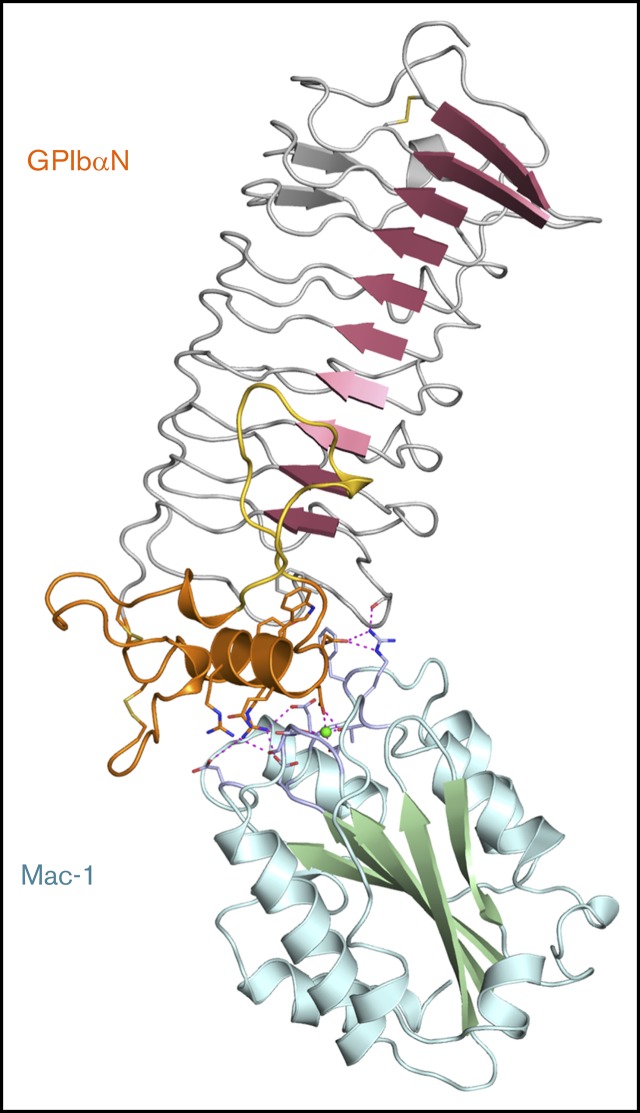
Cell-surface receptor interactions between leukocyte integrin macrophage-1 antigen (Mac-1, also known as CR3, αMβ2, CD11b/CD18) and platelet glycoprotein Ibα (GPIbα) are critical to vascular inflammation. To define the key residues at the binding interface, we used nuclear magnetic resonance (NMR) to assign the spectra of the mouse Mac-1 I-domain and mapped the residues contacting the mouse GPIbα N-terminal domain (GPIbαN) to the locality of the integrin metal ion-dependant adhesion site (MIDAS) surface. We next determined the crystal structures of the mouse GPIbαN and Mac-1 I-domain to 2 Å and 2.5 Å resolution, respectively. The mouse Mac-1 I-domain crystal structure reveals an active conformation that is stabilized by a crystal contact from the α7-helix with a glutamate side chain completing the octahedral coordination sphere of the MIDAS Mg ion. The amino acid sequence of the α7-helix and disposition of the glutamic acid matches the C-terminal capping region α-helix of GPIbα effectively acting as a ligand mimetic. Using these crystal structures in combination with NMR measurements and docking analysis, we developed a model whereby an acidic residue from the GPIbα leucine-rich repeat (LRR) capping α-helix coordinates directly to the Mac-1 MIDAS Mg ion. The Mac-1:GPIbαN complex involves additional interactions consolidated by an elongated pocket flanking the GPIbαN LRR capping α-helix. The GPIbαN α-helix has an HxxxE motif, which is equivalent by homology to RxxxD from the human GPIbαN. Subsequent mutagenesis of residues at this interface, coupled with surface plasmon resonance studies, confirmed the importance of GPIbαN residues H218, E222, and the Mac-1 MIDAS residue T209 to formation of the complex.
白细胞整合素巨噬细胞-1 抗原(Mac-1,也称为 CR3、αMβ2、CD11b/CD18)与血小板糖蛋白 Ibα(GPIbα)的细胞表面受体相互作用对于血管炎症至关重要。为了确定结合界面的关键残基,我们使用核磁共振(NMR)对小鼠 Mac-1 I 结构域进行了谱分配,并将与小鼠 GPIbαN 末端结构域(GPIbαN)接触的残基映射到整合素金属离子依赖性粘附位点(MIDAS)表面的位置。接下来,我们分别以 2 Å 和 2.5 Å 的分辨率确定了小鼠 GPIbαN 和 Mac-1 I 结构域的晶体结构。小鼠 Mac-1 I 结构域晶体结构揭示了一种活性构象,该构象由来自α7-螺旋的晶体接触稳定,谷氨酸侧链完成 MIDAS Mg 离子的八面体配位。α7-螺旋的氨基酸序列和谷氨酸的位置与 GPIbα 的 C 末端封端α-螺旋匹配,有效地充当配体模拟物。使用这些晶体结构结合 NMR 测量和对接分析,我们开发了一种模型,其中来自 GPIbα 亮氨酸丰富重复(LRR)封端α-螺旋的酸性残基直接与 Mac-1 MIDAS Mg 离子配位。Mac-1:GPIbαN 复合物涉及通过侧翼 GPIbαN LRR 封端α-螺旋的伸长口袋整合的其他相互作用。GPIbαN α-螺旋具有 HxxxE 基序,该基序通过同源性与人类 GPIbαN 的 RxxxD 等效。随后对该界面的残基进行突变,并结合表面等离子体共振研究,证实了 GPIbαN 残基 H218、E222 和 Mac-1 MIDAS 残基 T209 对复合物形成的重要性。