Shijith K P, Sood Munish, Sud Ajay Deep, Ghai Amresh
Department of Radiology, Army Hospital (R & R), Delhi Cantt, 110010, India.
Department of Orthopaedics, Command Hospital Chandimandir, Haryana, 134107, India.
Chin J Traumatol. 2019 Jun;22(3):177-181. doi: 10.1016/j.cjtee.2019.01.010. Epub 2019 Apr 13.
Glenoid bone defect and the defect on the posterior-superior surface of the humerus "Hill-Sachs lesion" are the commonly seen bony lesions in patients with recurrent dislocation shoulder. Computed tomography (CT) scan is considered as the best option in assessing the bony defects in the recurrent dislocation shoulder. The aim of this study was to assess the clinical and radiological co-relation in the patients with recurrent dislocation shoulder.
Forty-four patients of recurrent dislocation shoulder who were evaluated between January 2015 and December 2017 at a tertiary care center, clinically and radiologically using CT scan and meeting the inclusion criteria, were included. The correlation between the clinical history of the number of dislocations and the bone loss using CT scan was evaluated. Two sided statistical tests were performed at a significance level of α = 0.05. The analysis was conducted using IBM SPSS STATISTICS (version 22.0).
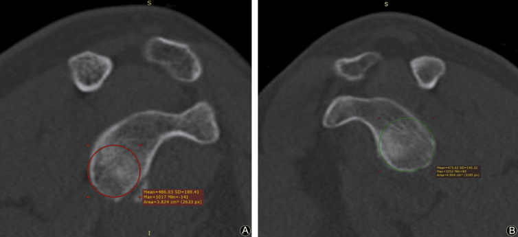
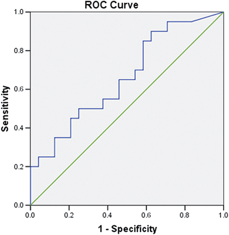
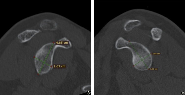
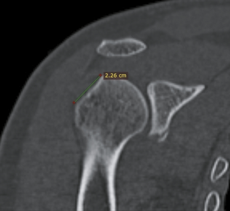
All the patients were male with mean age of 25.95 (SD ± 4.2) years were evaluated. Twenty-four patients sustained injury in sporting activities while 20 patients sustained injury in training. There were an average of 4.68 (SD ± 3.1, range 2-15, median 3) episodes of dislocation. Forty-one patients had the glenoid bone loss while 40 had the Hill-Sachs lesions. The mean glenoid width defect was 10.80% (range 0-27%) while the mean Hill-Sachs defect was 14.27 mm (range 0-26.6 mm). The mean area of bone loss of the glenoid surface was 10.81% (range 0-22.4%). The lesions were on track in 34 patients and off track in 10 patients.
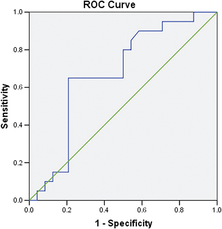
CT scan of the shoulder joint is an effective method for assessing the amount of bone loss. The number of dislocations are correlated significantly with off-track lesions and the amount of bone loss on the glenoid and Hill-Sachs lesion. The glenoid width bone loss of more than 9.80% or Hill-Sachs defect of more than 14.80 mm are the critical defects after which the frequency of dislocations increases.
盂骨缺损以及肱骨后上表面的缺损(“希尔-萨克斯损伤”)是复发性肩关节脱位患者中常见的骨损伤。计算机断层扫描(CT)被认为是评估复发性肩关节脱位骨缺损的最佳方法。本研究的目的是评估复发性肩关节脱位患者的临床与影像学相关性。
纳入2015年1月至2017年12月在一家三级医疗中心接受评估的44例复发性肩关节脱位患者,这些患者均符合纳入标准,接受了临床评估以及使用CT扫描的影像学评估。评估脱位次数的临床病史与使用CT扫描评估的骨质流失之间的相关性。进行双侧统计检验,显著性水平α = 0.05。使用IBM SPSS STATISTICS(版本22.0)进行分析。
所有患者均为男性,平均年龄25.95(标准差±4.2)岁。24例患者在体育活动中受伤,20例患者在训练中受伤。平均脱位次数为4.68(标准差±3.1,范围2 - 15,中位数3)次。41例患者有盂骨缺损,40例有希尔-萨克斯损伤。平均盂骨宽度缺损为10.80%(范围0 - 27%),而平均希尔-萨克斯缺损为14.27毫米(范围0 - 26.6毫米)。盂骨表面骨质流失的平均面积为10.81%(范围0 - 22.4%)。34例患者损伤情况良好,10例患者损伤情况不佳。
肩关节CT扫描是评估骨质流失量的有效方法。脱位次数与损伤情况不佳的损伤以及盂骨和希尔-萨克斯损伤处的骨质流失量显著相关。盂骨宽度骨质流失超过9.80%或希尔-萨克斯缺损超过14.80毫米是关键缺损,此后脱位频率会增加。