1 Stroke Unit Department of Neurology University Hospital of Nancy France.
3 Department of Neurology Stroke Center Hôpital Foch Suresnes France.
J Am Heart Assoc. 2019 May 21;8(10):e010962. doi: 10.1161/JAHA.118.010962.
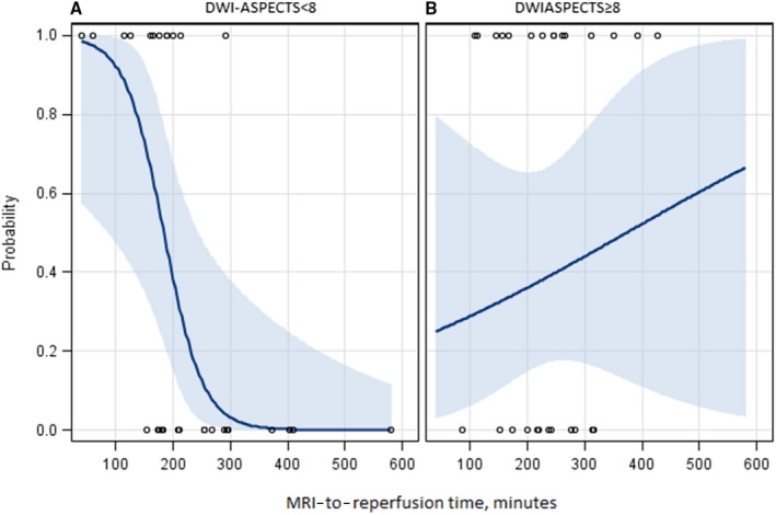
Background The association between time to reperfusion and clinical outcome is well known in anterior circulation strokes, whereas the impact of main time metrics remains unknown in posterior circulation strokes. We investigated the clinical effect of different time intervals from symptom onset to reperfusion on the 90-day clinical outcome in acute ischemic stroke patients with basilar artery occlusion, and especially in the subset population presenting a low stroke volume on baseline diffusion-weighted imaging. Methods and Results We studied patients included in the prospective, multicenter, observational ETIS (Endovascular Treatment in Ischemic Stroke) registry who had had basal artery occlusion and had achieved successful reperfusion (modified Thrombolysis In Cerebral Infarction 2b-3). Three time intervals (onset to reperfusion, onset to imaging, and imaging to reperfusion) were considered in all patients and separately in patients with pc- ASPECTS (posterior-circulation Alberta Stroke Program Early Computed Tomography Score) <8 and ≥8 on baseline diffusion-weighted imaging. The primary end point was good outcome defined as 90-day modified Rankin Scale scores of 0 to 2. Among the 95 included patients, 38 (40%) achieved a good outcome. In all patients, no significant association was found between the different time intervals and outcome. In patients evaluated with diffusion-weighted imaging (n=61) at baseline, a significant negative association was found between imaging-to-reperfusion time for patients with pc- ASPECTS <8 (adjusted odds ratio=0.4 per 30-minute increase; 95% CI 0.18-0.85; P=0.02) compared with those with pc- ASPECTS ≥8. Conclusions In patients with basilar artery occlusion and pc- ASPECTS <8 at baseline diffusion-weighted imaging, clinical outcome is highly dependent on the time from imaging to reperfusion, which suggests that rapid endovascular reperfusion should be performed after imaging in these patients.
在前循环卒中患者中,再灌注时间与临床结局之间的关联已得到充分证实,而在后循环卒中患者中,主要时间指标对临床结局的影响仍不清楚。我们研究了不同时间间隔(从症状发作到再灌注)对基底动脉闭塞的急性缺血性脑卒中患者 90 天临床结局的影响,特别是对基线弥散加权成像显示低卒中容积的亚组人群。
我们研究了前瞻性、多中心、观察性 ETIS(缺血性卒中血管内治疗)注册研究中纳入的基底动脉闭塞且成功再灌注的患者(改良脑梗死溶栓 2b-3)。在所有患者中考虑了三个时间间隔(从发作到再灌注、从发作到成像、从成像到再灌注),并分别在基线弥散加权成像上 pc-ASPECTS(后循环急性卒中治疗评分)<8 和≥8 的患者中进行了研究。主要终点是 90 天改良 Rankin 量表评分 0-2 的良好结局。在 95 例纳入的患者中,38 例(40%)达到了良好结局。在所有患者中,不同时间间隔与结局之间无显著相关性。在基线进行弥散加权成像评估的 61 例患者中,pc-ASPECTS<8 的患者,成像到再灌注时间与结局之间存在显著负相关(调整优势比为每 30 分钟增加 0.4;95%CI 0.18-0.85;P=0.02),而 pc-ASPECTS≥8 的患者则无相关性。
在基线弥散加权成像上 pc-ASPECTS<8 的基底动脉闭塞患者中,临床结局高度依赖于从成像到再灌注的时间,这表明这些患者应在成像后尽快进行血管内再灌注治疗。