Department of Epidemiology and Biostatistics, College of Public Health, Zhengzhou University, Zhengzhou 45001, China.
NHC Key Laboratory of Trace Elements and Endemic Diseases, Institute of Endemic Diseases, School of Public Health of Health Science Center, Xi'an Jiaotong University, Xi'an 710061, China.
Toxins (Basel). 2019 May 8;11(5):260. doi: 10.3390/toxins11050260.
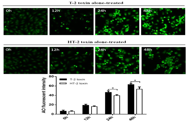
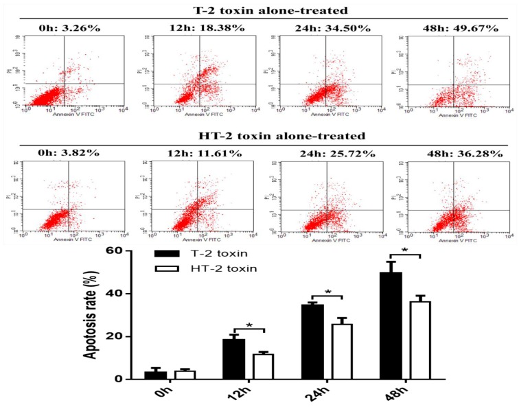
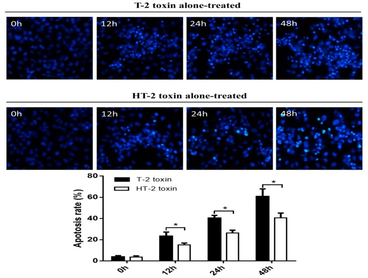
In this report, we have investigated the apoptosis and autophagy of chondrocytes induced by the T-2 and HT-2 toxins. The viability of chondrocytes was measured by the MTT assay. Malondialdehyde (MDA) and superoxide dismutase (SOD) kits were used to measure the oxidative stress of chondrocytes. The apoptosis of chondrocytes was measured using flow cytometry. Hoechst 33258 and MDC staining agents were introduced to analyze apoptosis and autophagy induction in chondrocytes, respectively. Protein expression of Bax, caspase-9, caspase-3, and Beclin1 was examined by western blotting analysis. The T-2 and HT-2 toxins significantly decreased the viability of chondrocytes in a time-dependent manner. The level of oxidative stress in chondrocytes induced by the T-2 toxin was significantly higher when compared with that of the HT-2 toxin. The apoptosis rate of chondrocytes induced by the T-2 toxin increased from 3.26 ± 1.03%, 18.38 ± 1.28%, 34.5 ± 1.40% to 49.67 ± 5.31%, whereas apoptosis rate of chondrocytes induced by the HT-2 toxin increased from 3.82 ± 1.03%, 11.61 ± 1.27%, 25.72 ± 2.95% to 36.28 ± 2.81% in 48 h incubation time. Hoechst 33258 staining confirmed that apoptosis of chondrocytes induced by the T-2 toxin was significantly higher than that observed when the chondrocytes were incubated with the HT-2 toxin. MDC staining revealed that the autophagy rate of chondrocytes induced by the T-2 toxin increased from 6.38% to 63.02%, whereas this rate induced by the HT-2 toxin changed from 6.08% to 53.33%. The expression levels of apoptosis and autophagy related proteins, Bax, caspase-9, caspase-3, and Beclin1 in chondrocytes induced by the T-2 toxin were significantly higher when compared with those levels induced by the HT-2 toxin.
在本报告中,我们研究了 T-2 和 HT-2 毒素诱导的软骨细胞凋亡和自噬。通过 MTT 测定法测量软骨细胞的活力。使用丙二醛 (MDA) 和超氧化物歧化酶 (SOD) 试剂盒测量软骨细胞的氧化应激。通过流式细胞术测量软骨细胞的凋亡。分别使用 Hoechst 33258 和 MDC 染色剂来分析软骨细胞的凋亡和自噬诱导。通过 Western 印迹分析检查 Bax、caspase-9、caspase-3 和 Beclin1 的蛋白表达。T-2 和 HT-2 毒素显著降低了软骨细胞的活力,呈时间依赖性。与 HT-2 毒素相比,T-2 毒素诱导的软骨细胞氧化应激水平显著升高。T-2 毒素诱导的软骨细胞凋亡率从 3.26±1.03%、18.38±1.28%、34.5±1.40%增加至 49.67±5.31%,而 HT-2 毒素诱导的软骨细胞凋亡率从 3.82±1.03%、11.61±1.27%、25.72±2.95%增加至 36.28±2.81%在 48 小时孵育时间内。Hoechst 33258 染色证实,T-2 毒素诱导的软骨细胞凋亡明显高于 HT-2 毒素孵育的软骨细胞。MDC 染色显示,T-2 毒素诱导的软骨细胞自噬率从 6.38%增加至 63.02%,而 HT-2 毒素诱导的自噬率从 6.08%增加至 53.33%。与 HT-2 毒素相比,T-2 毒素诱导的软骨细胞凋亡和自噬相关蛋白 Bax、caspase-9、caspase-3 和 Beclin1 的表达水平明显更高。