Department of Biochemistry and Molecular Biology, Beijing Normal University, Gene Engineering Drug and Biotechnology Beijing Key Laboratory, Beijing, China.
Biobank, The First Affiliated Hospital of Xi'an Jiaotong University, Xi'an, China.
Cancer Med. 2019 Jul;8(7):3553-3565. doi: 10.1002/cam4.2240. Epub 2019 May 15.
Patients with primary and metastatic brain cancer have an extremely poor prognosis, mostly due to the late diagnosis of disease. Urine, which lacks homeostatic mechanisms, is an ideal biomarker source that accumulates early and highly sensitive changes to provide information about the early stage of disease.
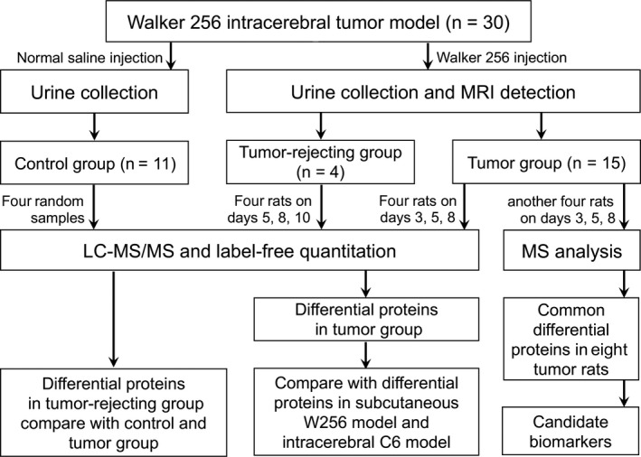
A rat model mimicking the local tumor growth process in the brain was established with intracerebral Walker 256 (W256) cell injection. Urine samples were collected on days 3, 5, and 8 after injection, and then analyzed by liquid chromatography coupled with tandem mass spectrometry.
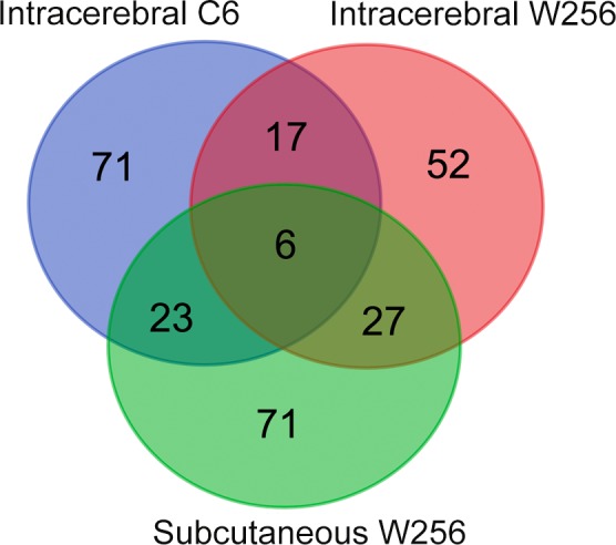
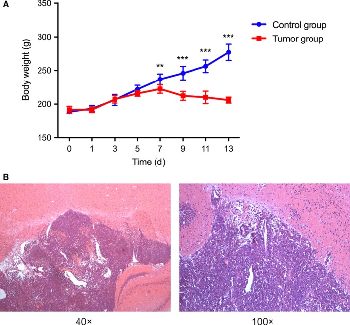
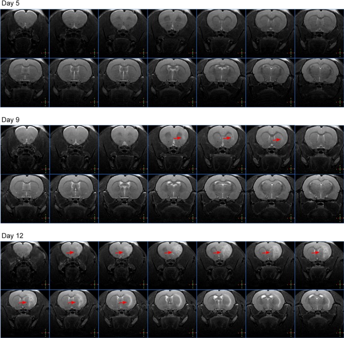
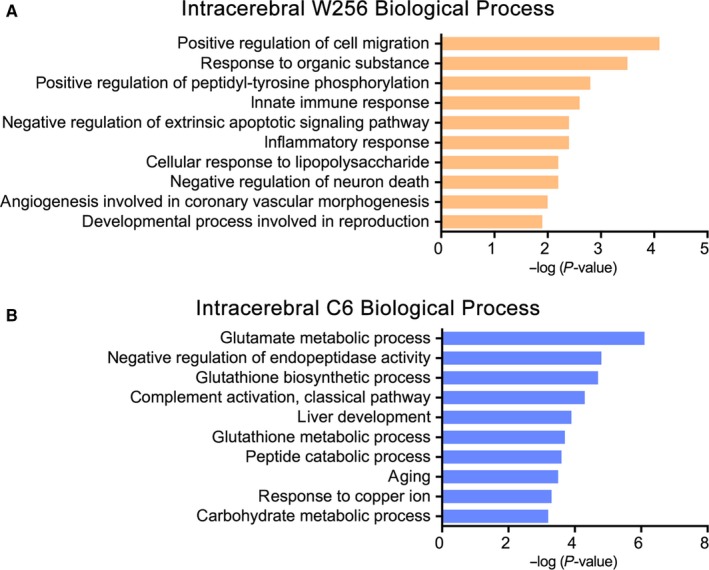
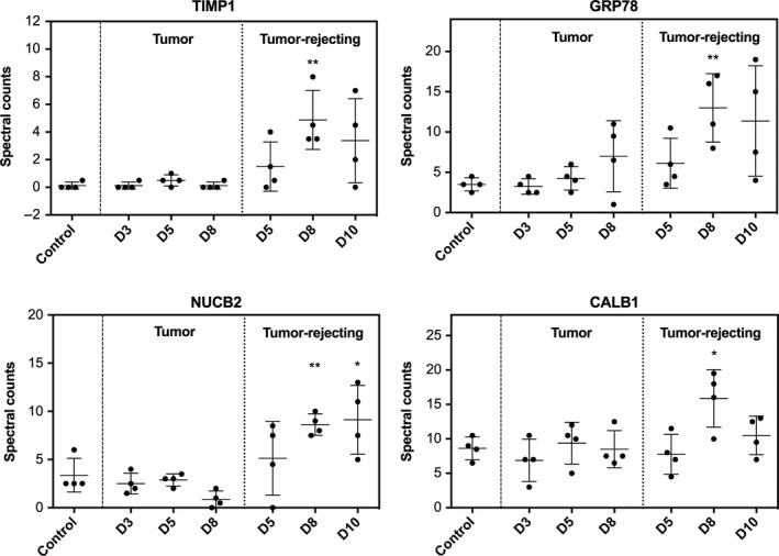
In the intracerebral W256 model, no obvious clinical manifestations or abnormal magnetic resonance imaging (MRI) signals were found on days 3 or 5; at these time points, 9 proteins were changed significantly in the urine of all eight tumor rats. On day 8, when tumors were detected by MRI, 25 differential proteins were identified, including 10 that have been reported to be closely related to brain metastasis or primary tumors. The differential urinary proteome was compared with those from the subcutaneous W256 model and the intracerebral C6 model. Few differential proteins overlapped, and specific differential protein patterns were observed among the three models.
These findings demonstrate that early changes in the urine proteome can be detected in the intracerebral W256 model. The urinary proteome can reflect the difference when tumor cells with different growth characteristics are inoculated into the brain and when identical tumor cells are inoculated into different areas, specifically, the subcutis and the brain.
原发性和转移性脑癌患者的预后极差,这主要是由于疾病的晚期诊断。尿液缺乏体内平衡机制,是一种理想的生物标志物来源,可积累早期且高度敏感的变化,提供有关疾病早期阶段的信息。
通过向脑内注射 Walker 256(W256)细胞建立了模拟脑内局部肿瘤生长过程的大鼠模型。在注射后第 3、5 和 8 天收集尿液样本,并通过液相色谱-串联质谱进行分析。
在颅内 W256 模型中,在第 3 天或第 5 天没有发现明显的临床症状或异常磁共振成像(MRI)信号;在这些时间点,所有 8 只肿瘤大鼠的尿液中有 9 种蛋白质发生了明显变化。在第 8 天,当 MRI 检测到肿瘤时,鉴定出 25 种差异蛋白,其中包括 10 种与脑转移或原发性肿瘤密切相关的蛋白。将差异尿蛋白质组与皮下 W256 模型和脑内 C6 模型进行比较。差异蛋白重叠很少,三个模型中观察到特定的差异蛋白模式。
这些发现表明,在颅内 W256 模型中可以检测到尿液蛋白质组的早期变化。尿液蛋白质组可以反映当具有不同生长特征的肿瘤细胞接种到脑内和当相同的肿瘤细胞接种到不同部位(即皮下和脑)时的差异。