Cardiology Unit, Cardiovascular Department, Fondazione IRCCS Casa Sollievo della Sofferenza, San Giovanni Rotondo (FG), Italy.
Pediatric Oncohematology Unit, Maternal and Child Developmental Age Department, Fondazione IRCCS Casa Sollievo della Sofferenza, San Giovanni Rotondo (FG), Italy.
Biomed Res Int. 2019 Apr 10;2019:2605323. doi: 10.1155/2019/2605323. eCollection 2019.
Pheochromocytoma is a rare neuroendocrine tumor, clinically characterized by high blood pressure, palpitations, and headache. It is often associated with abnormalities of the ventricular repolarization phase; the dispersion of ventricular repolarization is the basis for ventricular arrhythmias (torsion de point, ventricular tachycardia or ventricular fibrillation).
Analysis of abnormal ventricular repolarization focused on the presence and amount of U wave in patients affected by pheochromocytoma and its modification after surgery.
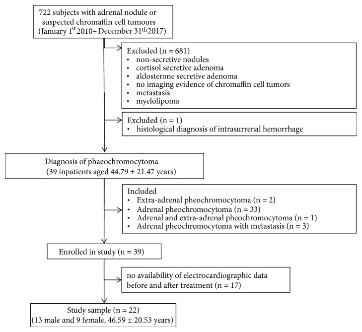
We reviewed pathology records of 722 patients admitted for adrenal nodule or suspected chromaffin-cell tumor and identified 39 patients affected by pheochromocytoma. Metanephrine, normetanephrine, and 3-methoxytyramine have been assessed by determining concentrations in 24-hour urine collection. Standard 12-lead electrocardiogram records have been reviewed with analysis of heart rate, P wave, PR interval, QRS duration, QTc, and U wave. Then we selected and compared 22 patients of 39 affected by pheochromocytoma, with both clinical and electrocardiographic data before and after surgery.
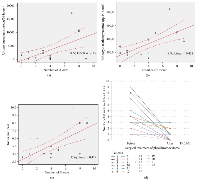
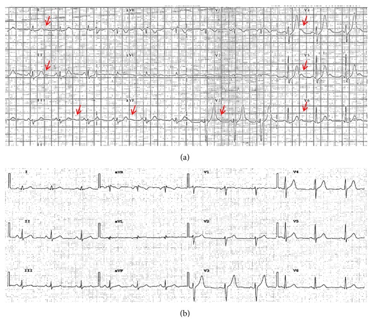
In our cohort of 39 patients affected by pheochromocytoma, we found U wave in ECG, before treatment, in 82.8 percent of patients, while only 37.0 percent after treatment (p<0.001) and we observed a statistically significant correlation between this wave and the urinary metanephrine. After surgery, in the selected 22 patients, we observed a clear significant reduction in systemic blood pressure, fasting glucose, metanephrine, normetanephrine, and 3-methoxytyramine. We found a significant reduction of U wave presence and leads involved in these patients after surgery (90.9% versus 9%). We observed a linear correlation between the amount of U waves in 12-lead electrocardiogram and metanephrine (r=0.333, p=0.015), 3-methoxytyramine levels (r=0.458, p=0.006), and tumor size (r=0.429, p=0.003).
In our retrospective analysis, patients affected by pheochromocytoma presented U wave in electrocardiogram. The presence and amount of U wave were associated with the metanephrine levels and the tumor size with significant reduction after surgical removal.
嗜铬细胞瘤是一种罕见的神经内分泌肿瘤,临床上以高血压、心悸和头痛为特征。它常伴有心室复极相的异常;心室复极离散是室性心律失常(扭转型室性心动过速、室性心动过速或心室颤动)的基础。
分析嗜铬细胞瘤患者异常心室复极的特点,重点观察 U 波的存在和数量,以及手术后的变化。
我们回顾性分析了 722 例因肾上腺结节或疑似嗜铬细胞瘤而入院的患者的病理记录,其中 39 例为嗜铬细胞瘤患者。通过测定 24 小时尿液收集物中的浓度,评估甲氧基去甲肾上腺素、去甲肾上腺素和 3-甲氧基酪胺。我们还回顾了标准 12 导联心电图记录,分析心率、P 波、PR 间期、QRS 持续时间、QTc 和 U 波。然后我们选择并比较了 39 例嗜铬细胞瘤患者中的 22 例,比较了他们术前和术后的临床和心电图数据。
在我们的 39 例嗜铬细胞瘤患者中,我们发现 82.8%的患者在治疗前的心电图上有 U 波,而只有 37.0%的患者在治疗后有 U 波(p<0.001),并且我们观察到这种波与尿中甲氧基去甲肾上腺素之间存在统计学显著相关性。在手术后的 22 例患者中,我们观察到全身血压、空腹血糖、甲氧基去甲肾上腺素、去甲肾上腺素和 3-甲氧基酪胺均显著降低。我们发现这些患者术后 U 波的存在和导联明显减少(90.9%对 9%)。我们观察到 12 导联心电图 U 波数量与甲氧基去甲肾上腺素(r=0.333,p=0.015)、3-甲氧基酪胺水平(r=0.458,p=0.006)和肿瘤大小(r=0.429,p=0.003)之间存在线性相关。
在我们的回顾性分析中,患有嗜铬细胞瘤的患者在心电图上出现 U 波。U 波的存在和数量与甲氧基去甲肾上腺素水平和肿瘤大小有关,手术切除后明显减少。