Department of Psychiatry and Psychology, Mayo Clinic, Rochester, Minnesota.
Temerty Centre for Therapeutic Brain Intervention, Centre for Addiction and Mental Health, Department of Psychiatry, University of Toronto, Ontario, Canada.
Int J Neuropsychopharmacol. 2019 Jul 1;22(7):435-444. doi: 10.1093/ijnp/pyz021.
The goal of this study was to examine baseline transcranial magnetic stimulation measures of cortical inhibition and excitability in depressed patients and characterize their longitudinal posttreatment changes.
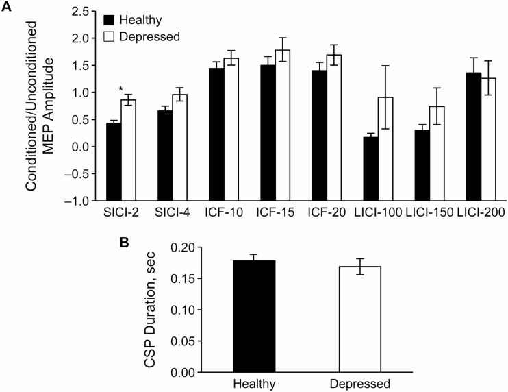
Fifteen adolescents (age 13-17 years) with moderate to severe major depressive disorder and 22 healthy controls (age 9-17) underwent single- and paired-pulse transcranial magnetic stimulation and clinical assessments. Transcranial magnetic stimulation measures included short-interval intracortical inhibition (2 and 4 milliseconds), long-interval intracortical inhibition (100, 150, and 200 milliseconds), cortical silent period, and intracortical facilitation (10, 15, and 20 milliseconds). Ten participants with major depressive disorder initiated antidepressant treatment or had dose adjustments. These participants were reassessed after treatment. Depression symptom severity was measured with the Children's Depression Rating Scale, Revised. Robust regression modeling compared healthy and depressed adolescents at baseline. Relationships between changes in cortical inhibition and changes in depressive symptom severity were assessed in the depressed adolescents receiving antidepressant treatment.
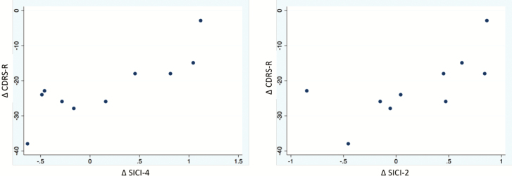
Our results revealed that at baseline, short-interval intracortical inhibition-2 was significantly reduced (Padj = .01) in depressed participants, suggesting impaired cortical inhibition compared with healthy controls. At follow-up, improvement in Children's Depression Rating Scale, Revised scores correlated with improvement in short-interval intracortical inhibition-4 amplitude (greater inhibition) after antidepressant treatment (R2 = 0.63; P = .01).
These results suggest that cortical inhibition measures may have promise as biomarkers in adolescents treated for depression.
本研究旨在检查抑郁患者的皮质抑制和兴奋性的基线经颅磁刺激测量值,并描述其治疗后的纵向变化。
15 名(13-17 岁)患有中重度重度抑郁症的青少年和 22 名健康对照者(9-17 岁)接受了单脉冲和双脉冲经颅磁刺激和临床评估。经颅磁刺激测量包括短间隔皮质内抑制(2 和 4 毫秒)、长间隔皮质内抑制(100、150 和 200 毫秒)、皮质静息期和皮质内易化(10、15 和 20 毫秒)。10 名患有重度抑郁症的参与者开始接受抗抑郁治疗或调整剂量。这些参与者在治疗后接受了重新评估。儿童抑郁评定量表修订版评估抑郁症状严重程度。稳健回归模型比较了健康和抑郁青少年的基线数据。在接受抗抑郁治疗的抑郁青少年中,评估了皮质抑制变化与抑郁症状严重程度变化之间的关系。
我们的结果表明,在基线时,短间隔皮质内抑制-2 明显降低(Padj=0.01),表明与健康对照组相比,皮质抑制受损。在随访时,儿童抑郁评定量表修订版评分的改善与抗抑郁治疗后短间隔皮质内抑制-4 振幅的改善(抑制作用增强)相关(R2=0.63;P=0.01)。
这些结果表明,皮质抑制测量值可能有希望作为接受抑郁症治疗的青少年的生物标志物。