Department of Pathology, Medical University of Vienna, Vienna, Austria.
Department of Virology, Medical University of Vienna, Vienna, Austria.
Liver Int. 2019 Oct;39(10):1876-1883. doi: 10.1111/liv.14137. Epub 2019 Jun 17.
Sporadic hepatitis E is an emerging indigenous disease in Europe induced by genotype 3 of the virus. While the disease takes an acute self-limited course in immunocompetent individuals, under immunocompromised conditions chronic hepatitis E might develop. The histology of chronic hepatitis E has not been described in detail systematically.
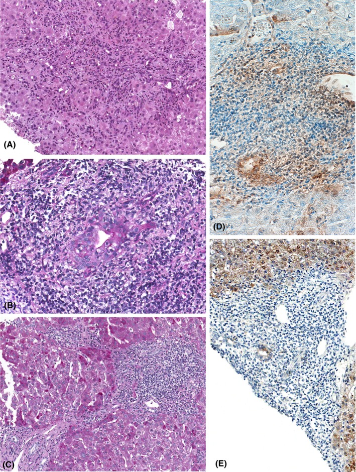
Liver biopsies from 19 immunosuppressed patients with chronic hepatitis E were collected: 17 were organ transplant recipients, one had a CD4-deficiency and one had received steroid therapy because of ulcerative colitis. Biopsies were processed with standard stains. Evaluation of histologic activity and fibrosis was performed according to Ishak. Additionally, immunohistochemistry with antibodies directed against open reading frame 2 and 3 of the virus was performed and liver biopsies were tested for hepatitis E virus RNA.
Biochemical data showed an increase in alanine transaminase, aspartate transaminase, gamma-glutamyl transferase and total bilirubin. Histopathology displayed typical features of chronic hepatitis with mild to moderate activity. The number of polymorphonuclear leucocytes was considerably increased and all patients had a florid cholangitis that presented as a destructive form in five of them. Hepatocytes and bile duct epithelia stained positive for hepatitis E virus by immunohistochemistry.
Chronic hepatitis E in immunocompromised individuals runs a similar course as hepatitis B and C and shows similar histopathology. However, the presence of destructive cholangitis in some cases accompanied by an increased number of polymorphonuclear leucocytes is markedly different. Immunohistochemically the virus is present in bile duct epithelia, seemingly the cause for cholangitis.
散发性戊型肝炎是欧洲新出现的一种由病毒基因型 3 引起的本土疾病。虽然在免疫功能正常的个体中,该病呈急性自限性病程,但在免疫功能低下的情况下,可能会发展为慢性戊型肝炎。慢性戊型肝炎的组织学尚未得到系统详细描述。
收集了 19 例免疫抑制患者的慢性戊型肝炎肝活检标本:17 例为器官移植受者,1 例为 CD4 缺陷,1 例因溃疡性结肠炎接受类固醇治疗。使用标准染色剂处理活检。根据 Ishak 标准评估组织学活动度和纤维化程度。此外,还进行了针对病毒开放阅读框 2 和 3 的免疫组织化学染色,并检测了肝活检中的戊型肝炎病毒 RNA。
生化数据显示丙氨酸转氨酶、天冬氨酸转氨酶、γ-谷氨酰转移酶和总胆红素升高。组织病理学显示出慢性肝炎的典型特征,活动度为轻度至中度。中性粒细胞数量显著增加,所有患者均有明显的胆管炎,其中 5 例呈破坏性形式。免疫组织化学染色显示肝细胞和胆管上皮呈戊型肝炎病毒阳性。
免疫抑制个体中的慢性戊型肝炎与乙型肝炎和丙型肝炎具有相似的病程,并显示出相似的组织病理学特征。然而,在某些情况下存在破坏性胆管炎伴中性粒细胞数量增加,这明显不同。免疫组织化学染色显示胆管上皮中有病毒存在,似乎是胆管炎的原因。