From the Department of Internal Medicine (Y.Y., Y.C., B.B., X.C., R.M.W., R.B.F., S.-G.W.), University of Iowa Carver College of Medicine.
Veterans Affairs Medical Center, Iowa City, IA (R.B.F.).
Hypertension. 2019 Jul;74(1):63-72. doi: 10.1161/HYPERTENSIONAHA.119.12651. Epub 2019 Jun 3.
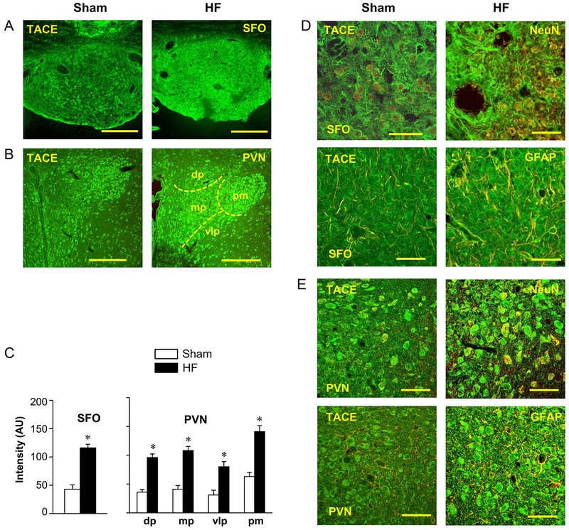
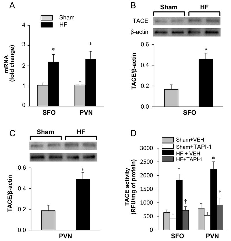
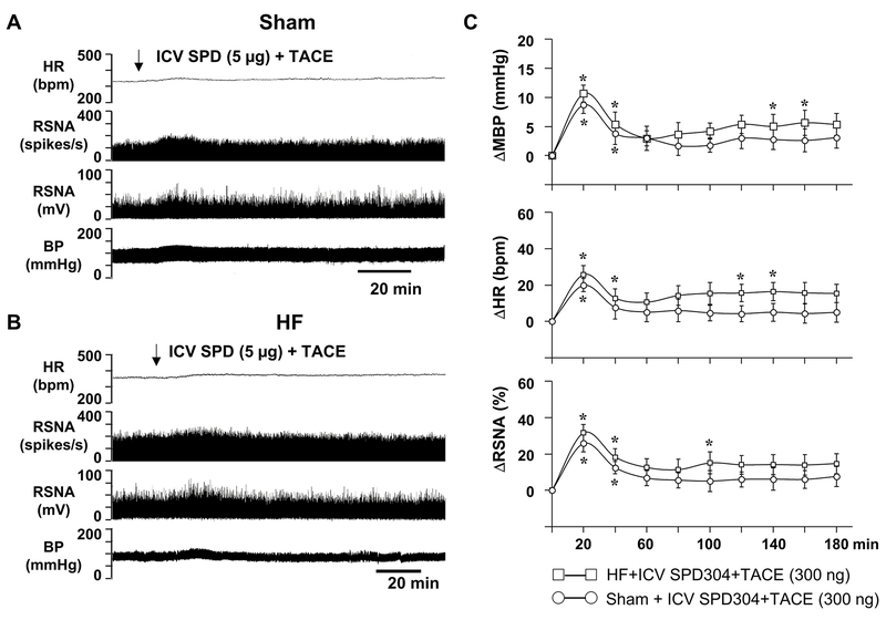
TNF-α (tumor necrosis factor-α) is initially synthesized as a transmembrane protein that is cleaved by TACE (TNF-α-converting enzyme) to release soluble TNF-α. The elevated level of TNF-α in the brain and circulation in heart failure (HF) suggests an increase in the TACE-mediated ectodomain shedding process. The present study sought to determine whether TACE is upregulated in cardiovascular/autonomic brain regions like subfornical organ and hypothalamic paraventricular nucleus in rats with ischemia-induced HF and whether TACE plays a role in TNF-α-driven sympathetic excitation. We found that TACE was expressed throughout the subfornical organ and paraventricular nucleus, with significantly higher levels in HF than in sham-operated (Sham) rats. Intracerebroventricular injection of recombinant TACE induced a mild increase in blood pressure, heart rate, and renal sympathetic nerve activity that peaked at 15 to 20 minutes in both Sham and HF rats. HF rats had a secondary prolonged increase in these variables that was prevented by the TNF-α inhibitor SPD304. Intracerebroventricular administration of the TACE inhibitor TNF-alpha protease inhibitor 1 decreased blood pressure, heart rate, and renal sympathetic nerve activity in Sham and HF rats, with an exaggerated reduction in heart rate and renal sympathetic nerve activity in the HF rats. Direct microinjection of TACE or TNF-alpha protease inhibitor 1 into paraventricular nucleus or subfornical organ of Sham and HF rats elicited blood pressure, heart rate, and renal sympathetic nerve activity responses similar to intracerebroventricular TACE or TNF-alpha protease inhibitor 1. Intracerebroventricular infusion of Ang II (angiotensin II) and IL (interleukin)-1β increased TACE expression in subfornical organ and paraventricular nucleus of normal rats. These data suggest that a TACE-mediated increase in soluble TNF-α in the brain contributes to sympathetic excitation in HF.
TNF-α(肿瘤坏死因子-α)最初作为一种跨膜蛋白合成,该蛋白被 TACE(TNF-α 转换酶)切割,从而释放可溶性 TNF-α。心力衰竭(HF)患者大脑和循环中 TNF-α 水平升高表明 TACE 介导的细胞外结构域脱落过程增加。本研究旨在确定 TACE 是否在缺血性 HF 大鼠的心血管/自主脑区(如穹窿下器官和下丘脑室旁核)中上调,以及 TACE 是否在 TNF-α 驱动的交感神经兴奋中发挥作用。我们发现 TACE 在整个穹窿下器官和室旁核中表达,HF 大鼠中的表达水平明显高于假手术(Sham)大鼠。重组 TACE 脑室内注射会引起 Sham 和 HF 大鼠的血压、心率和肾交感神经活动轻度增加,在 Sham 和 HF 大鼠中分别在 15 至 20 分钟达到峰值。HF 大鼠的这些变量出现继发性延长增加,这可被 TNF-α 抑制剂 SPD304 预防。脑室内给予 TACE 抑制剂 TNF-α 蛋白酶抑制剂 1 可降低 Sham 和 HF 大鼠的血压、心率和肾交感神经活动,HF 大鼠的心率和肾交感神经活动降低更为明显。TACE 或 TNF-α 蛋白酶抑制剂 1 直接微注射到 Sham 和 HF 大鼠的室旁核或穹窿下器官会引起类似于脑室内 TACE 或 TNF-α 蛋白酶抑制剂 1 的血压、心率和肾交感神经活动反应。脑室内输注 Ang II(血管紧张素 II)和 IL(白细胞介素)-1β 可增加正常大鼠穹窿下器官和室旁核中的 TACE 表达。这些数据表明,大脑中 TACE 介导的可溶性 TNF-α增加导致 HF 中的交感神经兴奋。