Schizophrenia Research Laboratory, Neuroscience Research Australia, Sydney, NSW, 2031, Australia.
School of Psychiatry, University of New South Wales, Sydney, NSW, 2052, Australia.
Mol Psychiatry. 2021 Mar;26(3):849-863. doi: 10.1038/s41380-019-0434-0. Epub 2019 Jun 5.
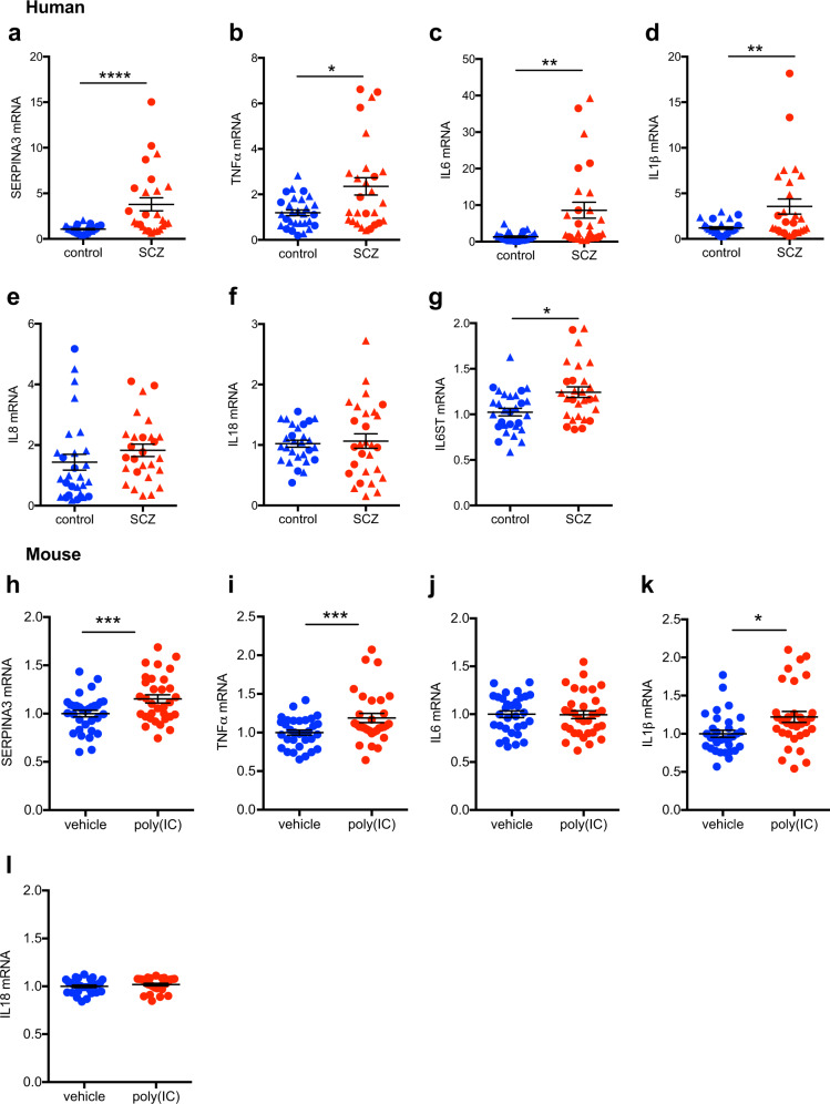
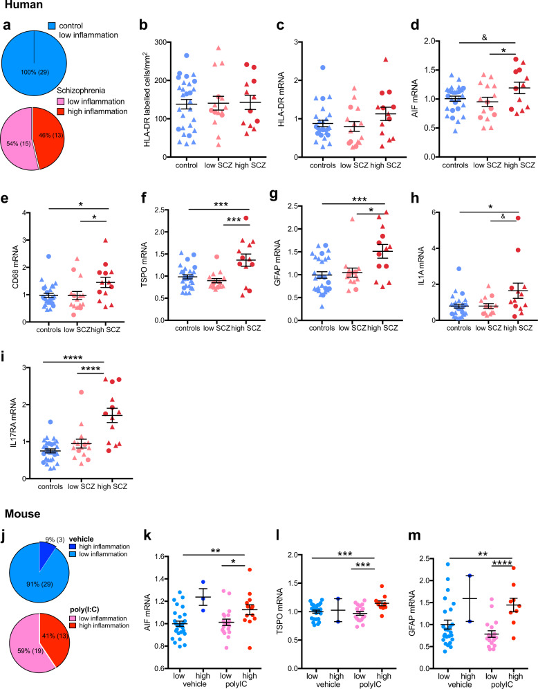
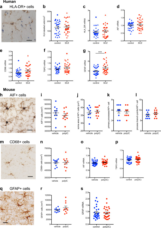
The pathophysiology of dopamine dysregulation in schizophrenia involves alterations at the ventral midbrain level. Given that inflammatory mediators such as cytokines influence the functional properties of midbrain dopamine neurons, midbrain inflammation may play a role in schizophrenia by contributing to presynaptic dopamine abnormalities. Thus, we quantified inflammatory markers in dopaminergic areas of the midbrain of people with schizophrenia and matched controls. We also measured these markers in midbrain of mice exposed to maternal immune activation (MIA) during pregnancy, an established risk factor for schizophrenia and other psychiatric disorders. We found diagnostic increases in SERPINA3, TNFα, IL1β, IL6, and IL6ST transcripts in schizophrenia compared with controls (p < 0.02-0.001). The diagnostic differences in these immune markers were accounted for by a subgroup of schizophrenia cases (~ 45%, 13/28) showing high immune status. Consistent with the human cohort, we identified increased expression of immune markers in the midbrain of adult MIA offspring (SERPINA3, TNFα, and IL1β mRNAs, all p ≤ 0.01), which was driven by a subset of MIA offspring (~ 40%, 13/32) with high immune status. There were no diagnostic (human cohort) or group-wise (mouse cohort) differences in cellular markers indexing the density and/or morphology of microglia or astrocytes, but an increase in the transcription of microglial and astrocytic markers in schizophrenia cases and MIA offspring with high inflammation. These data demonstrate that immune-related changes in schizophrenia extend to dopaminergic areas of the midbrain and exist in the absence of changes in microglial cell number, but with putative evidence of microglial and astrocytic activation in the high immune subgroup. MIA may be one of the contributing factors underlying persistent neuroimmune changes in the midbrain of people with schizophrenia.
精神分裂症中多巴胺失调的病理生理学涉及腹侧中脑水平的改变。鉴于细胞因子等炎症介质会影响中脑多巴胺神经元的功能特性,因此,中脑炎症可能通过导致突触前多巴胺异常而在精神分裂症中发挥作用。因此,我们对精神分裂症患者和匹配对照者的中脑多巴胺能区域的炎症标志物进行了定量检测。我们还在妊娠期间暴露于母体免疫激活 (MIA) 的小鼠的中脑中测量了这些标志物,MIA 是精神分裂症和其他精神障碍的既定风险因素。与对照组相比,我们发现精神分裂症患者的 SERPINA3、TNFα、IL1β、IL6 和 IL6ST 转录物的诊断增加(p < 0.02-0.001)。这些免疫标志物的诊断差异归因于具有高免疫状态的精神分裂症病例亚组(45%,13/28)。与人类队列一致,我们在成年 MIA 后代的中脑中发现了免疫标志物的表达增加(SERPINA3、TNFα 和 IL1β mRNAs,均 p ≤ 0.01),这是由具有高免疫状态的 MIA 后代亚组(40%,13/32)驱动的。在细胞标志物方面,没有诊断(人类队列)或组间(小鼠队列)差异,这些标志物可指示小胶质细胞或星形胶质细胞的密度和/或形态,但在具有高炎症的精神分裂症病例和 MIA 后代中,小胶质细胞和星形胶质细胞标志物的转录增加。这些数据表明,精神分裂症中的免疫相关变化扩展到中脑的多巴胺能区域,并且在小胶质细胞数量没有变化的情况下存在,但在高免疫亚组中存在小胶质细胞和星形胶质细胞激活的潜在证据。MIA 可能是精神分裂症患者中脑持续神经免疫变化的一个促成因素。