Schatten H, Walter M, Mazia D, Biessmann H, Paweletz N, Coffe G, Schatten G
Integrated Microscopy Resource for Biomedical Research, University of Wisconsin, Madison 53706.
Proc Natl Acad Sci U S A. 1987 Dec;84(23):8488-92. doi: 10.1073/pnas.84.23.8488.
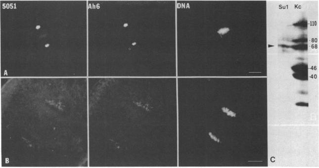
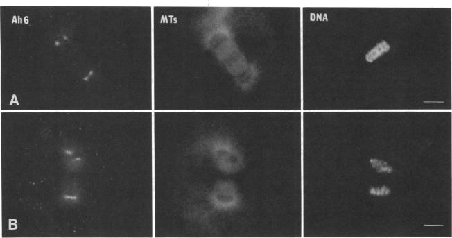
A mouse monoclonal antibody generated against Drosophila intermediate filament proteins (designated Ah6/5/9 and referred to herein as Ah6) is found to cross-react specifically with centrosomes in sea urchin eggs and with a 68-kDa antigen in eggs and isolated mitotic apparatus. When preparations stained with Ah6 are counterstained with a human autoimmune serum whose anti-centrosome activity has been established, the immunofluorescence images superimpose exactly. A more severe test of the specificity of the antibody demands that it display all of the stages of the centrosome cycle in the cell cycle: the flattening and spreading of the compact centrosomes followed by their division and the establishment of two compact poles. The test was made by an experimental design that uses a period of exposure of the eggs to 2-mercaptoethanol. This treatment allows observation of the stages of the centrosome cycle--separation, division, and bipolarization--while the chromosomes are arrested in metaphase. Mitosis is arrested in the presence of 0.1 M 2-mercaptoethanol. Chromosomes remain in a metaphase configuration while the centrosomes divide, producing four poles perpendicular to the original spindle axis. Microtubules are still present in the mitotic apparatus, as indicated by immunofluorescence and transmission electron microscopy. When 2-mercaptoethanol is removed, the chromosomes reorient to the poles of a tetrapolar (sometimes tripolar) mitotic apparatus. During the following cycle, the blastomeres form a monopolar mitotic apparatus. The observations of the centrosome cycle with the Ah6 antibody display very clearly all the stages that have been seen or deduced from work with other probes. The 68-kDa antigen that reacts with the Ah6 monoclonal antibody to Drosophila intermediate filament proteins must be a constant component of sea urchin centrosomes because it is present at all stages of the centrosome cycle.
一种针对果蝇中间丝蛋白产生的小鼠单克隆抗体(命名为Ah6/5/9,本文中称为Ah6),被发现能与海胆卵中的中心体以及卵和分离的有丝分裂器中的一种68 kDa抗原发生特异性交叉反应。当用Ah6染色的制剂用已确定具有抗中心体活性的人类自身免疫血清进行复染时,免疫荧光图像完全重叠。对该抗体特异性的更严格测试要求它能在细胞周期中显示中心体周期的所有阶段:紧密的中心体变平并扩散,随后进行分裂并形成两个紧密的极。该测试是通过一种实验设计进行的,该设计使用将卵暴露于2-巯基乙醇的一段时间。这种处理允许在染色体停滞在中期时观察中心体周期的各个阶段——分离、分裂和双极化。在0.1 M 2-巯基乙醇存在的情况下,有丝分裂被阻断。染色体保持中期构型,而中心体进行分裂,产生四个垂直于原始纺锤体轴的极。如免疫荧光和透射电子显微镜所示,微管仍存在于有丝分裂器中。当去除2-巯基乙醇时,染色体重新定向到四极(有时是三极)有丝分裂器的极。在随后的周期中,卵裂球形成单极有丝分裂器。用Ah6抗体对中心体周期的观察非常清楚地显示了所有已通过其他探针观察到或推断出的阶段。与针对果蝇中间丝蛋白的Ah6单克隆抗体发生反应的68 kDa抗原一定是海胆中心体的一个恒定成分,因为它在中心体周期的所有阶段都存在。