Max-Planck-Institute of Molecular Cell Biology and Genetics, Dresden, Germany.
Instutut de Biologie du Developpement de Marseille-Luminy, Aix-Marseille Universite, Marseille, France.
Traffic. 2019 Aug;20(8):601-617. doi: 10.1111/tra.12671.
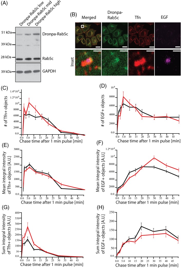
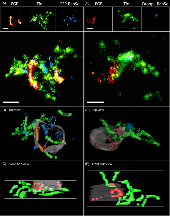
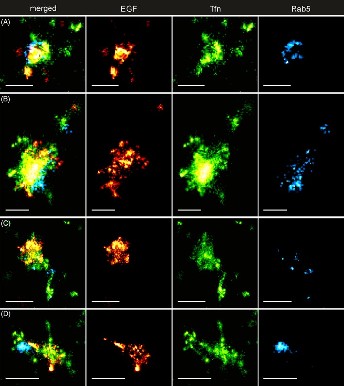
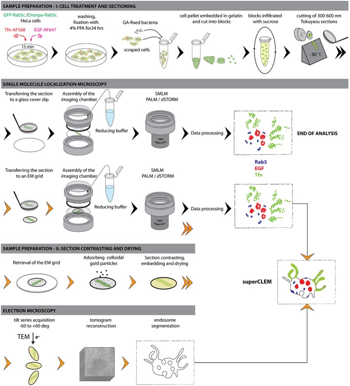
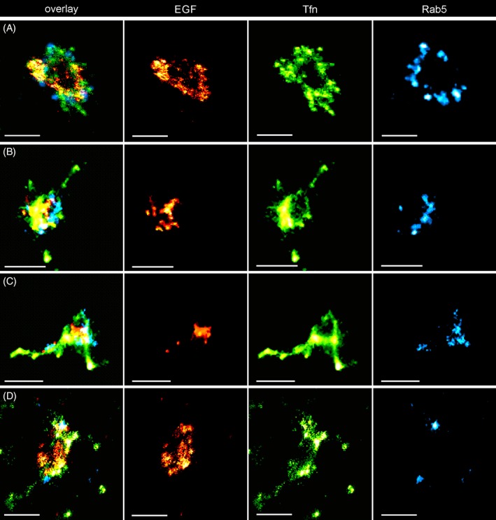
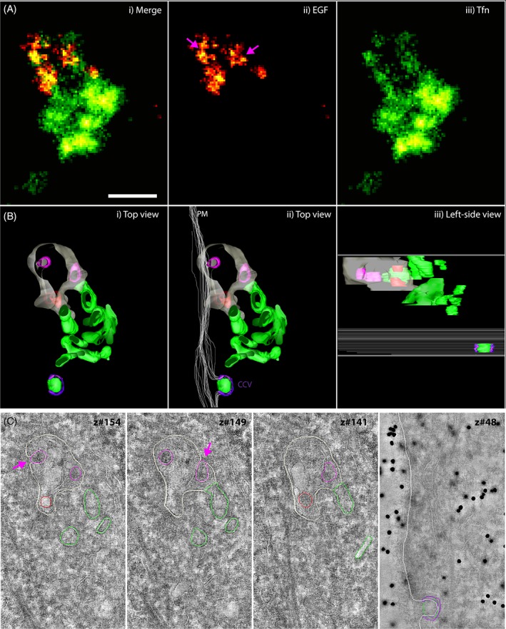
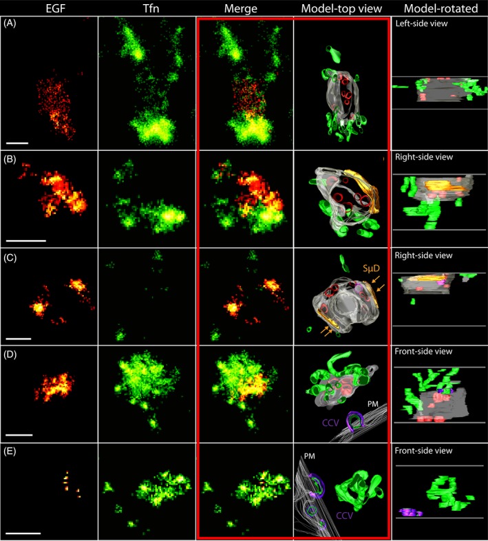
Many cellular organelles, including endosomes, show compartmentalization into distinct functional domains, which, however, cannot be resolved by diffraction-limited light microscopy. Single molecule localization microscopy (SMLM) offers nanoscale resolution but data interpretation is often inconclusive when the ultrastructural context is missing. Correlative light electron microscopy (CLEM) combining SMLM with electron microscopy (EM) enables correlation of functional subdomains of organelles in relation to their underlying ultrastructure at nanometer resolution. However, the specific demands for EM sample preparation and the requirements for fluorescent single-molecule photo-switching are opposed. Here, we developed a novel superCLEM workflow that combines triple-color SMLM (dSTORM & PALM) and electron tomography using semi-thin Tokuyasu thawed cryosections. We applied the superCLEM approach to directly visualize nanoscale compartmentalization of endosomes in HeLa cells. Internalized, fluorescently labeled Transferrin and EGF were resolved into morphologically distinct domains within the same endosome. We found that the small GTPase Rab5 is organized in nanodomains on the globular part of early endosomes. The simultaneous visualization of several proteins in functionally distinct endosomal sub-compartments demonstrates the potential of superCLEM to link the ultrastructure of organelles with their molecular organization at nanoscale resolution.
许多细胞细胞器,包括内体,显示出分隔成不同功能域的特征,但这无法通过受衍射极限限制的光学显微镜来分辨。单分子定位显微镜(SMLM)提供纳米级分辨率,但当缺少超微结构背景时,数据解释往往不明确。将 SMLM 与电子显微镜(EM)相结合的相关光电子显微镜(CLEM)能够在纳米分辨率下关联细胞器的功能亚区与其基础超微结构。然而,EM 样品制备的具体要求和荧光单分子光开关的要求是相互矛盾的。在这里,我们开发了一种新的超级 CLEM 工作流程,该流程结合了三色彩 SMLM(dSTORM 和 PALM)和使用半薄 Tokuyasu 解冻冷冻切片的电子断层扫描。我们应用超级 CLEM 方法直接可视化 HeLa 细胞中内体的纳米级分隔。内部化的、荧光标记的转铁蛋白和 EGF 在同一个内体中被分辨为形态上不同的结构域。我们发现,小分子 GTPase Rab5 在早期内体的球形部分上形成纳米结构域。在功能上不同的内体亚区中同时可视化几种蛋白质,证明了超级 CLEM 将细胞器的超微结构与其分子组织在纳米分辨率下关联的潜力。