Department of Immunology, Genetics and Pathology, Uppsala University, 75185 Uppsala, Sweden.
Molecular Immunology Laboratory, Shemyakin & Ovchinnikov Institute of Bioorganic Chemistry, Russian Academy of Sciences, 117997 Moscow, Russia.
Int J Mol Sci. 2019 Jun 21;20(12):3047. doi: 10.3390/ijms20123047.
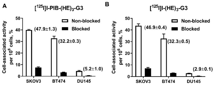
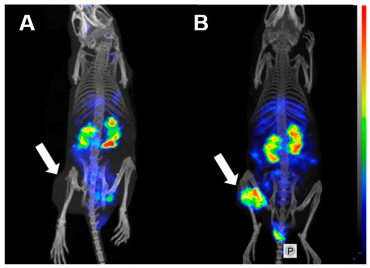
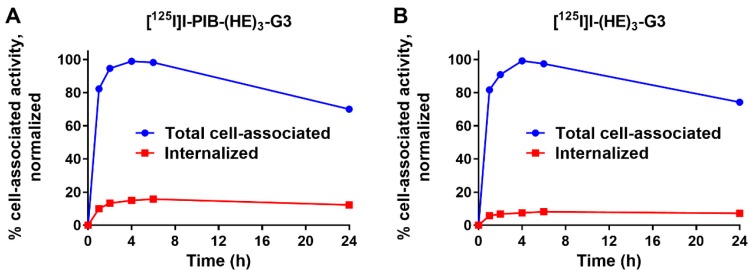
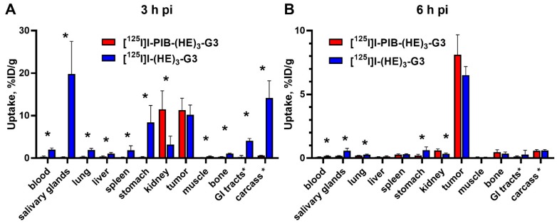
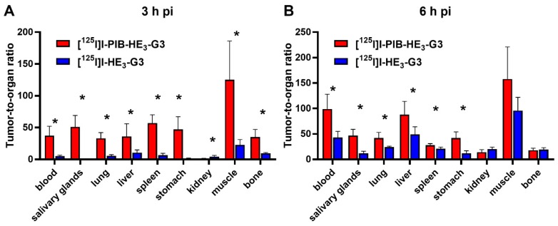
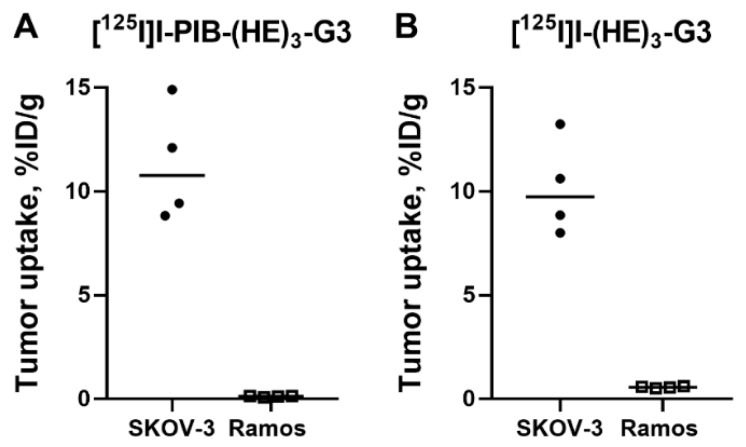
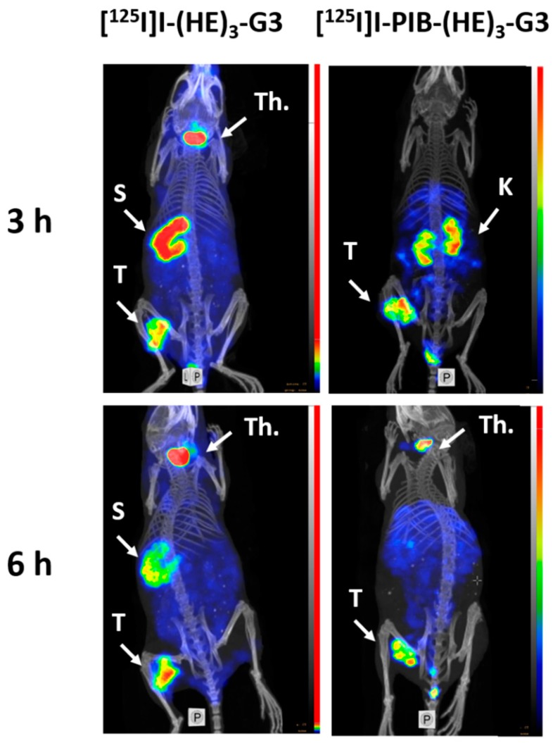
Radionuclide molecular imaging of human epidermal growth factor receptor 2 (HER2) in breast and gastroesophageal cancer might be used to stratify patients for HER2-targeted therapy as well as monitor treatment response and disease progression. Designed ankyrin repeat proteins (DARPins) are small engineered scaffold proteins with favorable properties for molecular imaging. Herein we compared two methods for labeling the anti-HER2 DARPin (HE)-G3, direct and indirect radioiodination. We hypothesized that the use of N-succinimidyl--iodobenzoate (SPIB) for radioiodination would facilitate the clearance of radiometabolites and improve the contrast of imaging. Both radiolabeled (HE)-G3 variants preserved their binding specificity and high affinity to HER2-expressing cells. The specificity of tumor targeting in vivo was also demonstrated. A biodistribution comparison of [I]I-(HE)-G3 and [I]I-PIB-(HE)-G3, in mice bearing HER2 expressing SKOV3 xenografts, showed rapid clearance of [I]I-PIB-(HE)-G3 from normal organs and tissues and low accumulation of activity in organs with NaI-symporter expression. Both radiolabeled (HE)-G3 variants had equal tumor uptake. Consequently, the indirect label provided higher tumor-to-blood and tumor-to-organ ratios compared with the direct label. Comparative Single Photon Emission Computed Tomography (SPECT)/CT imaging of HER2 expression in SKOV3 xenografts, using both radiolabeled DARPins, demonstrated the superior imaging contrast of the indirect label. Indirect radioiodination of (HE)-G3 using SPIB could be further applied for SPECT and PET imaging with iodine-123 and iodine-124.
用于乳腺癌和胃食管癌的人表皮生长因子受体 2 (HER2) 的放射性核素分子成像,可用于对 HER2 靶向治疗进行分层,以及监测治疗反应和疾病进展。设计的锚蛋白重复蛋白 (DARPins) 是具有有利分子成像特性的小型工程支架蛋白。本文比较了两种标记抗 HER2 DARPin (HE)-G3 的方法,直接和间接放射性碘标记。我们假设使用 N-琥珀酰亚胺基-4-碘苯甲酸酯 (SPIB) 进行放射性碘标记将有助于清除放射性代谢物并提高成像对比度。两种放射性标记的 (HE)-G3 变体都保留了其对 HER2 表达细胞的结合特异性和高亲和力。还在体内证明了肿瘤靶向的特异性。在表达 HER2 的 SKOV3 异种移植瘤小鼠中,对 [I]I-(HE)-G3 和 [I]I-PIB-(HE)-G3 的生物分布进行比较,显示 [I]I-PIB-(HE)-G3 从正常器官和组织中的快速清除以及在具有 NaI-转运蛋白表达的器官中活性的低积累。两种放射性标记的 (HE)-G3 变体的肿瘤摄取量相等。因此,间接标记物提供的肿瘤与血液和肿瘤与器官的比值均高于直接标记物。使用两种放射性标记的 DARPins 对 SKOV3 异种移植瘤中 HER2 表达进行单光子发射计算机断层扫描 (SPECT)/CT 比较成像,证明了间接标记物的成像对比度更好。使用 SPIB 对 (HE)-G3 进行间接放射性碘标记可进一步应用于碘-123 和碘-124 的 SPECT 和 PET 成像。