Czerwik Adriana, Olszewska Agnieszka, Starzomska Barbara, Korta Rafał, Henrich Manfred, Wrzosek Marcin, Schmidt Martin Jürgen
Department of Internal Medicine and Clinic of Horses, Dogs and Cats, Faculty of Veterinary Medicine, Wrocław University of Environmental and Life Sciences, pl. Grunwaldzki 47, 50-366, Wrocław, Poland.
Department of Veterinary Clinical Sciences, Small Animal Clinic, Justus-Liebig-University, Frankfurter Str. 108, 35392, Giessen, Germany.
Acta Vet Scand. 2019 Jun 25;61(1):32. doi: 10.1186/s13028-019-0467-z.
Multiple cartilaginous exostoses are a rare, benign, proliferative condition of cartilage and bone. They can be asymptomatic, or they may cause pain, lameness, paresis and even paralysis, depending on their location and size. In cases of spinal cord or nerve root compression, surgery is the treatment of choice. Therefore, an advanced imaging diagnostic work-up is indicated. Due to the unclear pathophysiology and progression of this condition, it is difficult to predict its prognosis.
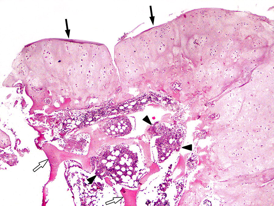
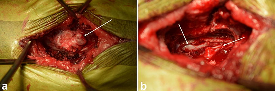
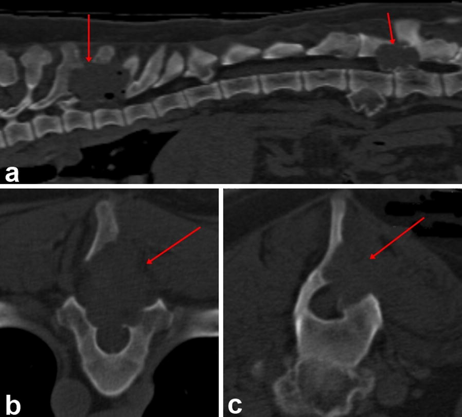
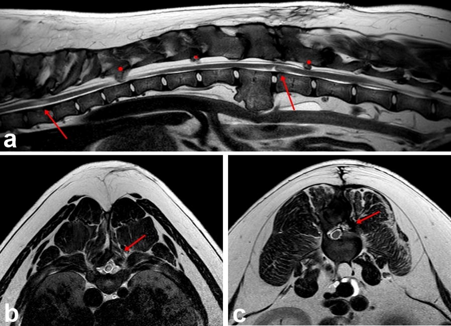
A 9-month-old female Swiss Mountain dog was presented with a history of gait abnormalities, kyphosis and hypersensitivity consistent with a thoracolumbar myelopathy. Multiple calcified masses, most prominent at the Th7-Th9 level and the L2-L3 level, were observed. Magnetic resonance imaging of the thoracolumbar vertebral column revealed severe dorsal spinal cord compressions near the dorsal arch of the Th7-Th9 and L2-L3 vertebrae. Two of these masses were removed surgically. The successful removal of both masses was confirmed by postoperative computed tomography. The histopathological examination of the resected tissue revealed multiple cartilaginous exostoses. The first neurological and magnetic resonance follow up examination carried out 6 months postoperatively showed improvement of the clinical status. At that time, no mass regrowth was observed. The last follow up neurological examination carried out 15 months postoperatively showed gait improvement and resolution of pain.
This is the first case report of multiple cartilaginous exostoses with a complete pre- and postoperative evaluation and a 15 month follow-up.
多发性软骨外生骨疣是一种罕见的、良性的软骨和骨增殖性疾病。它们可能无症状,也可能根据其位置和大小导致疼痛、跛行、轻瘫甚至瘫痪。在脊髓或神经根受压的情况下,手术是首选治疗方法。因此,需要进行先进的影像学诊断检查。由于这种疾病的病理生理学和进展尚不清楚,很难预测其预后。
一只9个月大的瑞士山地雌性犬,有步态异常、脊柱后凸和超敏反应病史,符合胸腰椎脊髓病。观察到多个钙化肿块,在胸7-胸9水平和腰2-腰3水平最为明显。胸腰椎脊柱的磁共振成像显示在胸7-胸9和腰2-腰3椎体背侧弓附近存在严重的脊髓背侧压迫。其中两个肿块通过手术切除。术后计算机断层扫描证实两个肿块均成功切除。对切除组织的组织病理学检查显示为多发性软骨外生骨疣。术后6个月进行的首次神经学和磁共振随访检查显示临床状况有所改善。当时,未观察到肿块复发。术后15个月进行的最后一次神经学随访检查显示步态改善且疼痛缓解。
这是第一例对多发性软骨外生骨疣进行完整术前和术后评估及15个月随访的病例报告。