• 文献检索
  • 文档翻译
  • 深度研究
  • 学术资讯
  • Suppr Zotero 插件Zotero 插件
  • 邀请有礼
  • 套餐&价格
  • 历史记录
应用&插件
Suppr Zotero 插件Zotero 插件浏览器插件Mac 客户端Windows 客户端微信小程序
定价
高级版会员购买积分包购买API积分包
服务
文献检索文档翻译深度研究API 文档MCP 服务
关于我们
关于 Suppr公司介绍联系我们用户协议隐私条款
关注我们

Suppr 超能文献

核心技术专利:CN118964589B侵权必究
粤ICP备2023148730 号-1Suppr @ 2026

文献检索

告别复杂PubMed语法,用中文像聊天一样搜索,搜遍4000万医学文献。AI智能推荐,让科研检索更轻松。

立即免费搜索

文件翻译

保留排版,准确专业,支持PDF/Word/PPT等文件格式,支持 12+语言互译。

免费翻译文档

深度研究

AI帮你快速写综述,25分钟生成高质量综述,智能提取关键信息,辅助科研写作。

立即免费体验

皮肤镜是诊断恶性雀斑样痣和恶性雀斑样痣黑色素瘤的好工具吗?一项荟萃分析。

Is dermoscopy a good tool for the diagnosis of lentigo maligna and lentigo maligna melanoma? A meta-analysis.

作者信息

Carapeba Murilo de Oliveira Lima, Alves Pineze Mariana, Nai Gisele Alborghetti

机构信息

Department of Dermatology, Universidade do Oeste Paulista (UNOESTE), Presidente Prudente, SP 19050-680, Brazil.

Medical School, Universidade do Oeste Paulista (UNOESTE), Presidente Prudente, SP, 19050-680, Brazil.

出版信息

Clin Cosmet Investig Dermatol. 2019 Jun 5;12:403-414. doi: 10.2147/CCID.S208717. eCollection 2019.

DOI:10.2147/CCID.S208717
PMID:31239744
原文链接:https://pmc.ncbi.nlm.nih.gov/articles/PMC6556474/
Abstract

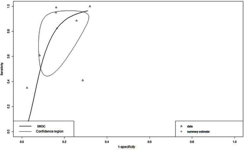
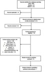
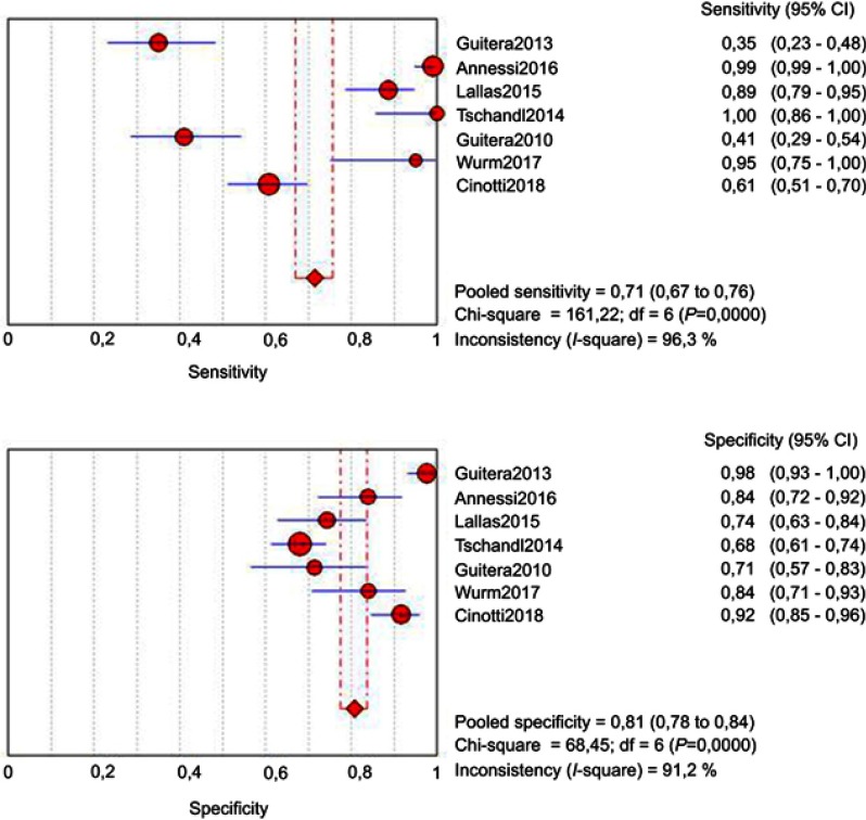
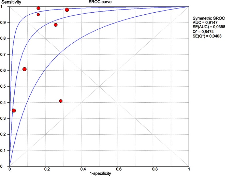
Dermoscopy is a low-cost examination performed by a dermatologist and good for the diagnosis of pigmented lesions. However, dermoscopy diagnosis of lentigo maligna (LM) and lentigo maligna melanoma (LMM) is still questionable. The objective of this study was to evaluate whether dermoscopy is an effective diagnostic method to diagnose LM/LMM from other pigmented skin lesions, and to identify which are the most frequent dermoscopic criteria associated with LM/LMM For this systematic review and meta-analysis, we used the following descriptors: dermoscopy, lentigo maligna, lentigo maligna melanoma, histopathology; and the following databases to search for articles: Cochrane Collaboration, MEDLINE; PMC (PubMed Central) - NIH (National Institutes of Health), EMBASE (The Excerpt Medical Database), and SCISEARCH, from inception to March 30, 2018. The evaluation of studies was performed using the QUADAS (Quality Assessment of Diagnostic Accuracy Studies)-2 tool. The PRISMA (Preferred Reporting Itens for Systematic Reviews and Meta-Analyses) and MOOSE (Meta-analysis Of Observational Studies in Epidemiology) guidelines were followed for data extraction. Also, we extracted from each study the dermoscopic criteria most commonly found in the lesions of LM/LMM. This systematic review included 15 articles for qualitative analysis (a total of 2,012 lesions evaluated) and 7 for meta-analysis. In the bivariate model the mean sensitivity was 0.824 and the mean specificity was 0.835. The area under the curve was 0.889. Rhomboid structures, pseudonetwork, and homogeneous areas were the most frequent dermoscopic criteria associated with LM/LMM. These findings suggest that dermoscopy has good accuracy in the diagnosis of LM/LMM.

摘要

皮肤镜检查是皮肤科医生进行的一种低成本检查,有助于色素性皮损的诊断。然而,皮肤镜对恶性雀斑样痣(LM)和恶性雀斑样痣黑色素瘤(LMM)的诊断仍存在疑问。本研究的目的是评估皮肤镜检查是否是一种从其他色素性皮肤损害中诊断LM/LMM的有效诊断方法,并确定与LM/LMM相关的最常见皮肤镜标准。对于这项系统评价和荟萃分析,我们使用了以下描述词:皮肤镜检查、恶性雀斑样痣、恶性雀斑样痣黑色素瘤、组织病理学;并使用以下数据库检索文章:Cochrane协作网、MEDLINE、PMC(PubMed Central)-NIH(美国国立卫生研究院)、EMBASE(医学文摘数据库)和SCISEARCH,检索时间从数据库建立至2018年3月30日。使用QUADAS(诊断准确性研究质量评估)-2工具对研究进行评估。数据提取遵循PRISMA(系统评价和荟萃分析优先报告条目)和MOOSE(流行病学观察性研究的荟萃分析)指南。此外,我们从每项研究中提取了LM/LMM皮损中最常见的皮肤镜标准。这项系统评价纳入了15篇文章进行定性分析(共评估2012个皮损),7篇文章进行荟萃分析。在双变量模型中,平均敏感性为0.824,平均特异性为0.835。曲线下面积为0.889。菱形结构、假网络和均匀区域是与LM/LMM相关的最常见皮肤镜标准。这些发现表明,皮肤镜检查在LM/LMM的诊断中具有良好的准确性。

https://cdn.ncbi.nlm.nih.gov/pmc/blobs/4802/6556474/b397cf9ddbfe/CCID-12-403-g0005.jpg
https://cdn.ncbi.nlm.nih.gov/pmc/blobs/4802/6556474/926658c4d347/CCID-12-403-g0001.jpg
https://cdn.ncbi.nlm.nih.gov/pmc/blobs/4802/6556474/7dbe586b39c2/CCID-12-403-g0002.jpg
https://cdn.ncbi.nlm.nih.gov/pmc/blobs/4802/6556474/4c9abac5fc1d/CCID-12-403-g0003.jpg
https://cdn.ncbi.nlm.nih.gov/pmc/blobs/4802/6556474/e3f7f2626b18/CCID-12-403-g0004.jpg
https://cdn.ncbi.nlm.nih.gov/pmc/blobs/4802/6556474/b397cf9ddbfe/CCID-12-403-g0005.jpg

相似文献

1
Is dermoscopy a good tool for the diagnosis of lentigo maligna and lentigo maligna melanoma? A meta-analysis.皮肤镜是诊断恶性雀斑样痣和恶性雀斑样痣黑色素瘤的好工具吗?一项荟萃分析。
Clin Cosmet Investig Dermatol. 2019 Jun 5;12:403-414. doi: 10.2147/CCID.S208717. eCollection 2019.
2
Lentigo Maligna: Keys to Dermoscopic Diagnosis.恶性雀斑样痣:皮肤镜诊断要点
Actas Dermosifiliogr. 2016 Jul-Aug;107(6):489-97. doi: 10.1016/j.ad.2016.01.001. Epub 2016 Feb 11.
3
Lentigo Maligna and Lentigo Maligna Melanoma of the External Ear: Clinical and Dermoscopic Features of 19 Patients.外耳恶性雀斑样痣和恶性雀斑样黑素瘤:19 例患者的临床和皮肤镜特征。
Exp Dermatol. 2024 Oct;33(10):e15188. doi: 10.1111/exd.15188.
4
A comparison of dermoscopic features among lentigo senilis/initial seborrheic keratosis, seborrheic keratosis, lentigo maligna and lentigo maligna melanoma on the face.面部脂溢性角化病/早期脂溢性角化病、脂溢性角化病、恶性雀斑样痣及恶性雀斑样痣黑色素瘤的皮肤镜特征比较
J Dermatol. 2004 Nov;31(11):884-9. doi: 10.1111/j.1346-8138.2004.tb00621.x.
5
Lentigo maligna and lentigo maligna melanoma in vivo differentiation with dermoscopy and reflectance confocal microscopy: A retrospective, multicentre study.利用皮肤镜和反射共聚焦显微镜对原位性恶性黑素细胞痣和恶性黑素瘤进行体内鉴别:一项回顾性、多中心研究。
J Eur Acad Dermatol Venereol. 2023 Nov;37(11):2293-2300. doi: 10.1111/jdv.19291. Epub 2023 Jul 21.
6
Reflectance confocal microscopy and dermoscopy aid in evaluating repigmentation within or adjacent to lentigo maligna melanoma surgical scars.反射式共聚焦显微镜和皮肤镜有助于评估黑素细胞痣恶性黑素瘤手术疤痕内或周围的复色情况。
J Eur Acad Dermatol Venereol. 2020 Jan;34(1):74-81. doi: 10.1111/jdv.15819. Epub 2019 Aug 23.
7
Amelanotic/hypomelanotic lentigo maligna: Dermoscopic and confocal features predicting diagnosis.无色素性/低色素性恶性雀斑样痣:预测诊断的皮肤镜和共聚焦特征。
J Eur Acad Dermatol Venereol. 2023 Feb;37(2):303-310. doi: 10.1111/jdv.18636. Epub 2022 Oct 17.
8
Clinical findings are not helpful in detecting lentigo maligna melanoma in patients with biopsy-proven lentigo maligna.临床发现对于检测已确诊为良性恶性黑素瘤的患者的良性恶性黑素瘤并无帮助。
J Eur Acad Dermatol Venereol. 2022 Dec;36(12):2325-2330. doi: 10.1111/jdv.18346. Epub 2022 Jul 8.
9
Dermoscopy vs. reflectance confocal microscopy for the diagnosis of lentigo maligna.皮肤镜检查与反射共聚焦显微镜检查在诊断恶性雀斑样痣中的对比。
J Eur Acad Dermatol Venereol. 2018 Aug;32(8):1284-1291. doi: 10.1111/jdv.14791. Epub 2018 Feb 6.
10
Reviewing Challenges in the Diagnosis and Treatment of Lentigo Maligna and Lentigo-Maligna Melanoma.审视恶性雀斑样痣及恶性雀斑样痣黑色素瘤诊断与治疗中的挑战。
Rare Cancers Ther. 2015;3(1):133-145. doi: 10.1007/s40487-015-0012-9. Epub 2015 Oct 15.

引用本文的文献

1
Machine Learning-based Complementary Artificial Intelligence Model for Dermoscopic Diagnosis of Pigmented Skin Lesions in Resource-limited Settings.基于机器学习的辅助人工智能模型用于资源有限环境下色素性皮肤病变的皮肤镜诊断
Plast Reconstr Surg Glob Open. 2025 Jul 28;13(7):e7004. doi: 10.1097/GOX.0000000000007004. eCollection 2025 Jul.
2
Characteristics of patients with melanoma with non‑melanoma skin cancer comorbidity: Practical implications based on a retrospective study.合并非黑色素瘤皮肤癌的黑色素瘤患者的特征:基于一项回顾性研究的实际意义。
Oncol Lett. 2025 Mar 4;29(5):214. doi: 10.3892/ol.2025.14960. eCollection 2025 May.
3

本文引用的文献

1
Dermoscopy vs. reflectance confocal microscopy for the diagnosis of lentigo maligna.皮肤镜检查与反射共聚焦显微镜检查在诊断恶性雀斑样痣中的对比。
J Eur Acad Dermatol Venereol. 2018 Aug;32(8):1284-1291. doi: 10.1111/jdv.14791. Epub 2018 Feb 6.
2
Histopathologic and Immunohistochemical Correlates of Confocal Descriptors in Pigmented Facial Macules on Photodamaged Skin.光损伤皮肤色素沉着性面部斑疹共聚焦描述符的组织病理学和免疫组织化学相关性
JAMA Dermatol. 2017 Aug 1;153(8):771-780. doi: 10.1001/jamadermatol.2017.1323.
3
Correlation between digital epiluminescence microscopy parameters and histopathological changes in lentigo maligna and solar lentigo: A dermoscopic index for the diagnosis of lentigo maligna.
Skin Cancer Diagnosis by Lesion, Physician, and Examination Type: A Systematic Review and Meta-Analysis.
基于病变、医生及检查类型的皮肤癌诊断:一项系统评价与Meta分析
JAMA Dermatol. 2025 Feb 1;161(2):135-146. doi: 10.1001/jamadermatol.2024.4382.
4
Cutaneous Melanoma: An Overview of Physiological and Therapeutic Aspects and Biotechnological Use of Serine Protease Inhibitors.皮肤黑色素瘤:丝氨酸蛋白酶抑制剂的生理和治疗方面概述及生物技术应用。
Molecules. 2024 Aug 16;29(16):3891. doi: 10.3390/molecules29163891.
5
Perifollicular linear projections: A dermatoscopic criterion for the diagnosis of lentigo maligna on the face.毛囊周线性突起:面部恶性雀斑样痣诊断的皮肤镜标准
J Am Acad Dermatol. 2024 Jan;90(1):52-57. doi: 10.1016/j.jaad.2023.07.1036. Epub 2023 Aug 25.
6
Line-field confocal optical coherence tomography of lentigo maligna with horizontal and vertical histopathologic correlations.水平和垂直组织病理学相关性的恶性雀斑样痣线阵共聚焦光学相干断层扫描。
J Cutan Pathol. 2023 Feb;50(2):118-122. doi: 10.1111/cup.14321. Epub 2022 Sep 19.
7
Dermoscopy of external ear melanoma (EEM).耳部黑色素瘤的皮肤镜检查(EEM)。
Arch Dermatol Res. 2023 Jul;315(5):1381-1387. doi: 10.1007/s00403-022-02342-2. Epub 2022 Mar 17.
8
Hyperspectral Imaging Reveals Spectral Differences and Can Distinguish Malignant Melanoma from Pigmented Basal Cell Carcinomas: A Pilot Study.高光谱成像揭示光谱差异并能区分恶性黑色素瘤与色素性基底细胞癌:一项初步研究。
Acta Derm Venereol. 2021 Feb 19;101(2):adv00405. doi: 10.2340/00015555-3755.
数字表皮透光显微镜参数与恶性黑素细胞痣和日光性黑子的组织病理学变化之间的相关性:恶性黑素细胞痣诊断的一种皮肤镜指标。
J Am Acad Dermatol. 2017 Feb;76(2):234-243. doi: 10.1016/j.jaad.2016.08.032. Epub 2016 Oct 26.
4
Diagnostic accuracy of reflectance confocal microscopy using VivaScope for detecting and monitoring skin lesions: a systematic review.使用VivaScope反射共聚焦显微镜检测和监测皮肤病变的诊断准确性:一项系统评价
Clin Exp Dermatol. 2017 Apr;42(3):266-275. doi: 10.1111/ced.13055. Epub 2017 Feb 20.
5
The value of reflectance confocal microscopy in diagnosis of flat pigmented facial lesions: a prospective study.反射式共聚焦显微镜在诊断扁平色素性面部病变中的价值:一项前瞻性研究。
J Eur Acad Dermatol Venereol. 2017 Aug;31(8):1349-1354. doi: 10.1111/jdv.14171. Epub 2017 May 15.
6
Measures of Diagnostic Accuracy: Basic Definitions.诊断准确性的测量:基本定义。
EJIFCC. 2009 Jan 20;19(4):203-11. eCollection 2009 Jan.
7
Reviewing Challenges in the Diagnosis and Treatment of Lentigo Maligna and Lentigo-Maligna Melanoma.审视恶性雀斑样痣及恶性雀斑样痣黑色素瘤诊断与治疗中的挑战。
Rare Cancers Ther. 2015;3(1):133-145. doi: 10.1007/s40487-015-0012-9. Epub 2015 Oct 15.
8
Dermoscopic clues to differentiate facial lentigo maligna from pigmented actinic keratosis.面部恶性雀斑样痣与色素性光化性角化病的皮肤镜线索鉴别。
Br J Dermatol. 2016 May;174(5):1079-85. doi: 10.1111/bjd.14355. Epub 2016 Jan 20.
9
Diagnosis and management of lentigo maligna: a review.恶性雀斑样痣的诊断与管理:综述
Drugs Context. 2015 May 29;4:212281. doi: 10.7573/dic.212281. eCollection 2015.
10
Clinical and dermoscopic characteristics of melanomas on nonfacial chronically sun-damaged skin.非面部慢性日光损伤皮肤黑素瘤的临床和皮肤镜特征。
J Am Acad Dermatol. 2015 Jun;72(6):1027-35. doi: 10.1016/j.jaad.2015.02.1117. Epub 2015 Mar 29.