School of Electronic Engineering, College of IT Engineering, Kyungpook National University, Daegu, South Korea.
Department of Ophthalmology, School of Medicine, Kyungpook National University, Daegu, South Korea.
J Biophotonics. 2019 Nov;12(11):e201900098. doi: 10.1002/jbio.201900098. Epub 2019 Jul 23.
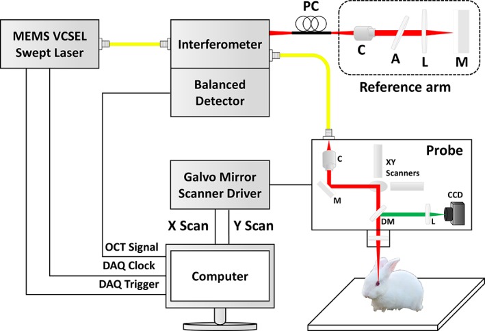
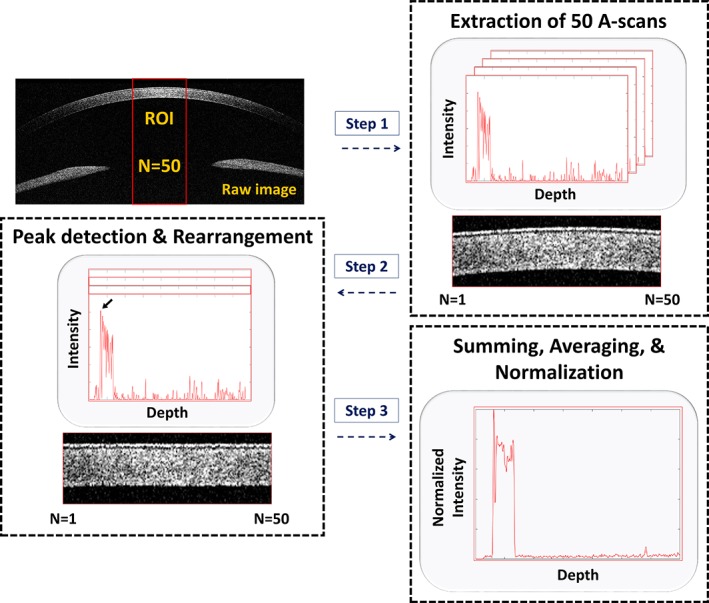
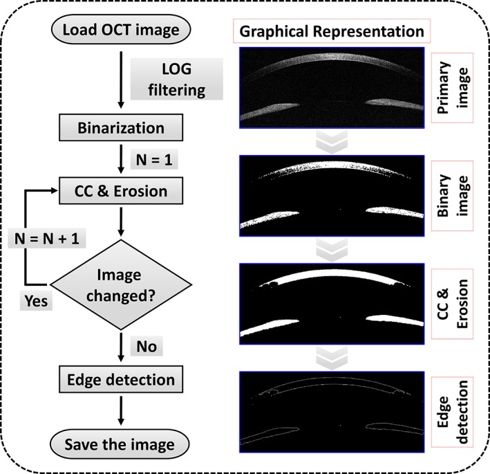
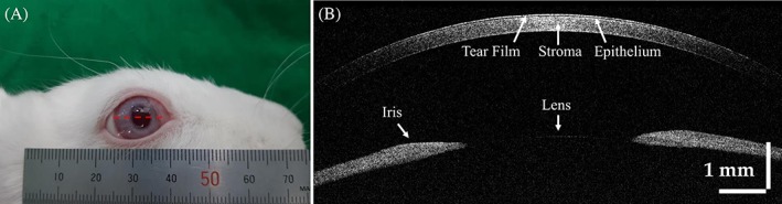
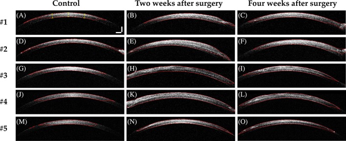
Corneal transplantation by full-thickness penetrating keratoplasty with human donor tissue is a widely accepted treatment for damaged or diseased corneas. Although corneal transplantation has a high success rate, a shortage of high-quality donor tissue is a considerable limitation. Therefore, bioengineered corneas could be an effective solution for this limitation, and a decellularized extracellular matrix comprises a promising scaffold for their fabrication. In this study, three-dimensional bioprinted decellularized collagen sheets were implanted into the stromal layer of the cornea of five rabbits. We performed in vivo noninvasive monitoring of the rabbit corneas using swept-source optical coherence tomography (OCT) after implanting the collagen sheets. Anterior segment OCT images and averaged amplitude-scans were acquired biweekly to monitor corneal thickness after implantation for 1 month. The averaged cornea thickness in the control images was 430.3 ± 5.9 μm, while the averaged thickness after corneal implantation was 598.5 ± 11.8 μm and 564.5 ± 12.5 μm at 2 and 4 weeks, respectively. The corneal thickness reduction of 34 μm confirmed the biocompatibility through the image analysis of the depth-intensity profile base. Moreover, hematoxylin and eosin staining supported the biocompatibility evaluation of the bioprinted decellularized collagen sheet implantation. Hence, the developed bioprinted decellularized collagen sheets could become an alternative solution to human corneal donor tissue, and the proposed image analysis procedure could be beneficial to confirm the success of the surgery.
全层穿透性角膜移植术用人体供体组织进行角膜移植是一种广泛接受的治疗受损或患病角膜的方法。尽管角膜移植的成功率很高,但高质量供体组织的短缺是一个相当大的限制。因此,生物工程角膜可能是解决这一限制的有效方法,而去细胞化的细胞外基质是制造它们的有前途的支架。在这项研究中,将三维生物打印的去细胞胶原片植入五只兔子的角膜基质层。我们在植入胶原片后使用扫频源光学相干断层扫描(OCT)对兔子角膜进行了体内非侵入性监测。在前节 OCT 图像和平均幅度扫描中,每两周采集一次,以监测植入后 1 个月的角膜厚度。对照图像中角膜的平均厚度为 430.3±5.9μm,而植入后 2 和 4 周的平均厚度分别为 598.5±11.8μm和 564.5±12.5μm。通过深度-强度剖面图的图像分析,角膜厚度减少 34μm 证实了生物相容性。此外,苏木精-伊红染色支持生物打印的去细胞胶原片植入的生物相容性评估。因此,开发的生物打印的去细胞胶原片可以成为人类角膜供体组织的替代方案,并且所提出的图像分析程序可能有助于确认手术的成功。