• 文献检索
  • 文档翻译
  • 深度研究
  • 学术资讯
  • Suppr Zotero 插件Zotero 插件
  • 邀请有礼
  • 套餐&价格
  • 历史记录
应用&插件
Suppr Zotero 插件Zotero 插件浏览器插件Mac 客户端Windows 客户端微信小程序
定价
高级版会员购买积分包购买API积分包
服务
文献检索文档翻译深度研究API 文档MCP 服务
关于我们
关于 Suppr公司介绍联系我们用户协议隐私条款
关注我们

Suppr 超能文献

核心技术专利:CN118964589B侵权必究
粤ICP备2023148730 号-1Suppr @ 2026

文献检索

告别复杂PubMed语法,用中文像聊天一样搜索,搜遍4000万医学文献。AI智能推荐,让科研检索更轻松。

立即免费搜索

文件翻译

保留排版,准确专业,支持PDF/Word/PPT等文件格式,支持 12+语言互译。

免费翻译文档

深度研究

AI帮你快速写综述,25分钟生成高质量综述,智能提取关键信息,辅助科研写作。

立即免费体验

碳-羟基-麻黄碱正电子发射断层显像/计算机断层扫描在嗜铬细胞瘤和副神经节瘤诊断中的应用

C-hydroxy-ephedrine-PET/CT in the Diagnosis of Pheochromocytoma and Paraganglioma.

作者信息

Vyakaranam Achyut Ram, Crona Joakim, Norlén Olov, Hellman Per, Sundin Anders

机构信息

Section of Radiology & Molecular Imaging, Department of Surgical Sciences, Uppsala University, Akademiska Sjukhuset, SE-751 85 Uppsala, Sweden.

Department of Surgical Sciences, Uppsala University, Akademiska Sjukhuset, SE-751 85 Uppsala, Sweden.

出版信息

Cancers (Basel). 2019 Jun 19;11(6):847. doi: 10.3390/cancers11060847.

DOI:10.3390/cancers11060847
PMID:31248124
原文链接:https://pmc.ncbi.nlm.nih.gov/articles/PMC6627429/
Abstract

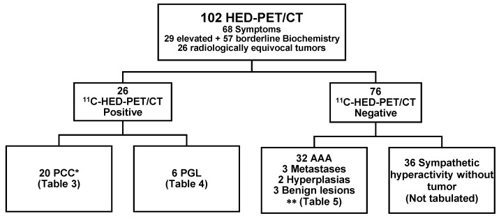
Pheochromocytomas (PCC) and paragangliomas (PGL) may be difficult to diagnose because of vague and uncharacteristic symptoms and equivocal biochemical and radiological findings. This was a retrospective cohort study in 102 patients undergoing C-hydroxy-ephedrine (C-HED)-PET/CT because of symptoms and/or biochemistry suspicious for PCC/PGL and/or with radiologically equivocal adrenal incidentalomas. Correlations utilized CT/MRI, clinical, biochemical, surgical, histopathological and follow-up data. C-HED-PET/CT correctly identified 19 patients with PCC and six with PGL, missed one PCC, attained one false positive result (nodular hyperplasia) and correctly excluded PCC/PGL in 75 patients. Sensitivity, specificity, positive and negative predictive values of C-HED-PET/CT for PCC/PGL diagnosis was 96%, 99%, 96% and 99%, respectively. In 41 patients who underwent surgical resection and for whom correlation to histopathology was available, the corresponding figures were 96%, 93%, 96% and 93%, respectively. Tumor C-HED-uptake measurements (standardized uptake value, tumor-to-normal-adrenal ratio) were unrelated to symptoms of catecholamine excess ( > 0.05) and to systolic blood pressure ( > 0.05). In PCC/PGL patients, norepinephrine and systolic blood pressure increased in parallel ( = 0.22, = 0.016). C-HED-PET/CT was found to be an accurate tool to diagnose and rule out PCC/PGL in complex clinical scenarios and for the characterization of equivocal adrenal incidentalomas. PET measurements of tumor C-HED uptake were not helpful for tumor characterization.

摘要

嗜铬细胞瘤(PCC)和副神经节瘤(PGL)可能难以诊断,因为其症状模糊且不具特征性,生化和放射学检查结果也不明确。这是一项回顾性队列研究,研究对象为102例因症状和/或生化检查结果怀疑患有PCC/PGL和/或肾上腺意外瘤放射学表现不明确而接受间碘苄胍(C-HED)-PET/CT检查的患者。研究采用了CT/MRI、临床、生化、手术、组织病理学和随访数据。C-HED-PET/CT正确识别出19例PCC患者和6例PGL患者,漏诊1例PCC,出现1例假阳性结果(结节性增生),并正确排除了75例患者的PCC/PGL。C-HED-PET/CT诊断PCC/PGL的敏感性、特异性、阳性预测值和阴性预测值分别为96%、99%、96%和99%。在41例接受手术切除且可与组织病理学进行对比的患者中,相应数值分别为96%、93%、96%和93%。肿瘤C-HED摄取测量值(标准化摄取值、肿瘤与正常肾上腺比值)与儿茶酚胺过量症状(>0.05)和收缩压(>0.05)无关。在PCC/PGL患者中,去甲肾上腺素和收缩压呈平行升高(r = 0.22,P = 0.016)。研究发现,C-HED-PET/CT是在复杂临床情况下诊断和排除PCC/PGL以及明确肾上腺意外瘤特征的准确工具。肿瘤C-HED摄取的PET测量值对肿瘤特征的判断并无帮助。

https://cdn.ncbi.nlm.nih.gov/pmc/blobs/f8ba/6627429/7cbbefe707a7/cancers-11-00847-g004.jpg
https://cdn.ncbi.nlm.nih.gov/pmc/blobs/f8ba/6627429/90d517fb2a53/cancers-11-00847-g001.jpg
https://cdn.ncbi.nlm.nih.gov/pmc/blobs/f8ba/6627429/46e25b37a3b6/cancers-11-00847-g002.jpg
https://cdn.ncbi.nlm.nih.gov/pmc/blobs/f8ba/6627429/87091df6e174/cancers-11-00847-g003.jpg
https://cdn.ncbi.nlm.nih.gov/pmc/blobs/f8ba/6627429/7cbbefe707a7/cancers-11-00847-g004.jpg

相似文献

1
C-hydroxy-ephedrine-PET/CT in the Diagnosis of Pheochromocytoma and Paraganglioma.碳-羟基-麻黄碱正电子发射断层显像/计算机断层扫描在嗜铬细胞瘤和副神经节瘤诊断中的应用
Cancers (Basel). 2019 Jun 19;11(6):847. doi: 10.3390/cancers11060847.
2
Localization and prediction of malignant potential in recurrent pheochromocytoma/paraganglioma (PCC/PGL) using 18F-FDG PET/CT.利用18F-FDG PET/CT对复发性嗜铬细胞瘤/副神经节瘤(PCC/PGL)进行恶性潜能的定位及预测
Acta Radiol. 2014 Jun;55(5):631-40. doi: 10.1177/0284185113504330. Epub 2013 Sep 14.
3
Genetic status determines F-FDG uptake in pheochromocytoma/paraganglioma.基因状态决定嗜铬细胞瘤/副神经节瘤中F-FDG的摄取。
J Med Imaging Radiat Oncol. 2017 Dec;61(6):745-752. doi: 10.1111/1754-9485.12620. Epub 2017 Jun 5.
4
Pheochromocytomas: detection with 11C hydroxyephedrine PET.嗜铬细胞瘤:用11C - 羟基麻黄碱PET进行检测
Radiology. 2004 Feb;230(2):423-8. doi: 10.1148/radiol.2302021678.
5
Adrenocortical Tumors and Pheochromocytoma/Paraganglioma Initially Mistaken as Neuroblastoma-Experiences From the GPOH-MET Registry.肾上腺皮质肿瘤和嗜铬细胞瘤/副神经节瘤最初误诊为神经母细胞瘤——来自 GPOH-MET 注册中心的经验。
Front Endocrinol (Lausanne). 2022 Jun 17;13:918435. doi: 10.3389/fendo.2022.918435. eCollection 2022.
6
(11)C-hydroxyephedrine positron emission tomography in the postoperative management of pheochromocytoma and paraganglioma.(11)C-羟基麻黄碱正电子发射断层扫描在嗜铬细胞瘤和副神经节瘤术后管理中的应用
Neuroendocrinology. 2014;100(1):60-70. doi: 10.1159/000365516. Epub 2014 Jul 5.
7
11C-hydroxyephedrine positron emission tomography imaging of pheochromocytoma: a single center experience over 11 years.11C-羟基麻黄碱正电子发射断层扫描成像在嗜铬细胞瘤中的应用:单中心 11 年经验。
J Clin Endocrinol Metab. 2012 Jul;97(7):2423-32. doi: 10.1210/jc.2011-3342. Epub 2012 May 4.
8
(68)Ga-DOTATATE and (18)F-FDG PET/CT in Paraganglioma and Pheochromocytoma: utility, patterns and heterogeneity.(68)镓-多柔比星和(18)氟-脱氧葡萄糖PET/CT在副神经节瘤和嗜铬细胞瘤中的应用:效用、表现形式及异质性
Cancer Imaging. 2016 Aug 17;16(1):22. doi: 10.1186/s40644-016-0084-2.
9
Surgical outcome of laparoscopic surgery, including laparoendoscopic single-site surgery, for retroperitoneal paraganglioma compared with adrenal pheochromocytoma.腹腔镜手术(包括经腹腔镜单部位手术)治疗腹膜后副神经节瘤与肾上腺嗜铬细胞瘤的手术结果比较。
J Endourol. 2014 Jun;28(6):686-92. doi: 10.1089/end.2013.0706. Epub 2014 Mar 24.
10
Whole-body PET/CT with 11C-meta-hydroxyephedrine in tumors of the sympathetic nervous system: feasibility study and comparison with 123I-MIBG SPECT/CT.11C-间羟基麻黄碱全身PET/CT在交感神经系统肿瘤中的应用:可行性研究及与123I-MIBG SPECT/CT的比较
J Nucl Med. 2006 Oct;47(10):1635-42.

引用本文的文献

1
Non-invasive imaging of sympathetic innervation of the pancreas in individuals with type 2 diabetes.2 型糖尿病患者胰腺交感神经支配的无创成像。
Diabetologia. 2024 Jan;67(1):199-208. doi: 10.1007/s00125-023-06039-7. Epub 2023 Nov 7.
2
Management of pheochromocytomas and paragangliomas: Review of current diagnosis and treatment options.处理嗜铬细胞瘤和副神经节瘤:当前诊断和治疗选择的综述。
Cancer Med. 2023 Jul;12(13):13942-13957. doi: 10.1002/cam4.6010. Epub 2023 May 5.
3
Primary adrenal tuberculosis infection in patients with Behcet's disease presenting as isolated adrenal metastasis by F-FDG PET/CT: a rare case report and literature review.

本文引用的文献

1
Performance of Ga-DOTA-Conjugated Somatostatin Receptor-Targeting Peptide PET in Detection of Pheochromocytoma and Paraganglioma: A Systematic Review and Metaanalysis.镓-DOTA 偶联生长抑素受体靶向肽 PET 在嗜铬细胞瘤和副神经节瘤检测中的性能:系统评价和荟萃分析。
J Nucl Med. 2019 Mar;60(3):369-376. doi: 10.2967/jnumed.118.211706. Epub 2018 Jul 20.
2
18F-FDOPA PET/CT Uptake Parameters Correlate with Catecholamine Secretion in Human Pheochromocytomas.18F-FDOPA PET/CT 摄取参数与人类嗜铬细胞瘤儿茶酚胺分泌相关。
Neuroendocrinology. 2018;107(3):228-236. doi: 10.1159/000491578. Epub 2018 Jun 27.
3
Role of DOTATATE-PET/CT in preoperative assessment of phaeochromocytoma and paragangliomas.
白塞病患者原发性肾上腺结核感染表现为F-FDG PET/CT显示的孤立性肾上腺转移:1例罕见病例报告及文献复习
Gland Surg. 2021 Dec;10(12):3431-3442. doi: 10.21037/gs-21-511.
4
Diagnosis for Pheochromocytoma and Paraganglioma: A Joint Position Statement of the Korean Pheochromocytoma and Paraganglioma Task Force.嗜铬细胞瘤和副神经节瘤的诊断:韩国嗜铬细胞瘤和副神经节瘤工作组的联合立场声明。
Endocrinol Metab (Seoul). 2021 Apr;36(2):322-338. doi: 10.3803/EnM.2020.908. Epub 2021 Apr 6.
5
Metastatic Pheochromocytomas and Abdominal Paragangliomas.转移性嗜铬细胞瘤和腹部副神经节瘤
J Clin Endocrinol Metab. 2021 Apr 23;106(5):e1937-e1952. doi: 10.1210/clinem/dgaa982.
6
COVID-19: Virology, biology and novel laboratory diagnosis.COVID-19:病毒学、生物学和新型实验室诊断。
J Gene Med. 2021 Feb;23(2):e3303. doi: 10.1002/jgm.3303. Epub 2021 Jan 6.
7
Recent advances in radiotracers targeting norepinephrine transporter: structural development and radiolabeling improvements.近年来靶向去甲肾上腺素转运体的放射性示踪剂的研究进展:结构发展和放射性标记的改进。
J Neural Transm (Vienna). 2020 Jun;127(6):851-873. doi: 10.1007/s00702-020-02180-4. Epub 2020 Apr 9.
8
Pheochromocytoma (PHEO) and Paraganglioma (PGL).嗜铬细胞瘤(PHEO)和副神经节瘤(PGL)。
Cancers (Basel). 2019 Sep 18;11(9):1391. doi: 10.3390/cancers11091391.
DOTATATE-PET/CT在嗜铬细胞瘤和副神经节瘤术前评估中的作用。
Clin Endocrinol (Oxf). 2018 Aug;89(2):139-147. doi: 10.1111/cen.13737. Epub 2018 May 24.
4
Superiority of Ga-DOTATATE over F-FDG and anatomic imaging in the detection of succinate dehydrogenase mutation (SDHx )-related pheochromocytoma and paraganglioma in the pediatric population.镓-DOTATATE 比 F-FDG 和解剖成像在儿童人群中检测琥珀酸脱氢酶突变(SDHx)相关嗜铬细胞瘤和副神经节瘤方面更具优势。
Eur J Nucl Med Mol Imaging. 2018 May;45(5):787-797. doi: 10.1007/s00259-017-3896-9. Epub 2017 Dec 4.
5
Quantitative F-DOPA PET/CT in pheochromocytoma: the relationship between tumor secretion and its biochemical phenotype.定量 F-DOPA PET/CT 在嗜铬细胞瘤中的应用:肿瘤分泌与其生化表型的关系。
Eur J Nucl Med Mol Imaging. 2018 Feb;45(2):278-282. doi: 10.1007/s00259-017-3833-y. Epub 2017 Sep 16.
6
Follow-up of patients with adrenal incidentaloma, in accordance with the European society of endocrinology guidelines: Could we be safe?按照欧洲内分泌学会指南对肾上腺偶发瘤患者进行随访:我们能放心吗?
J Endocrinol Invest. 2017 Mar;40(3):331-333. doi: 10.1007/s40618-016-0558-x. Epub 2016 Oct 15.
7
PET/CT comparing (68)Ga-DOTATATE and other radiopharmaceuticals and in comparison with CT/MRI for the localization of sporadic metastatic pheochromocytoma and paraganglioma.PET/CT比较(68)镓-多柔比星和其他放射性药物,并与CT/MRI比较用于散发性转移性嗜铬细胞瘤和副神经节瘤的定位。
Eur J Nucl Med Mol Imaging. 2016 Sep;43(10):1784-91. doi: 10.1007/s00259-016-3357-x. Epub 2016 Mar 21.
8
Prospective comparison of (68)Ga-DOTATATE and (18)F-FDOPA PET/CT in patients with various pheochromocytomas and paragangliomas with emphasis on sporadic cases.(68)镓- DOTATATE与(18)氟- FDOPA PET/CT在各类嗜铬细胞瘤和副神经节瘤患者中的前瞻性比较,重点关注散发病例。
Eur J Nucl Med Mol Imaging. 2016 Jul;43(7):1248-57. doi: 10.1007/s00259-015-3268-2. Epub 2015 Dec 5.
9
Superiority of [68Ga]-DOTATATE PET/CT to Other Functional Imaging Modalities in the Localization of SDHB-Associated Metastatic Pheochromocytoma and Paraganglioma.[68Ga]-DOTATATE PET/CT在SDHB相关转移性嗜铬细胞瘤和副神经节瘤定位方面相对于其他功能成像模态的优越性。
Clin Cancer Res. 2015 Sep 1;21(17):3888-95. doi: 10.1158/1078-0432.CCR-14-2751. Epub 2015 Apr 14.
10
The incremental benefit of functional imaging in pheochromocytoma/paraganglioma: a systematic review.功能成像在嗜铬细胞瘤/副神经节瘤中的增量效益:一项系统评价。
Endocrine. 2015 Sep;50(1):176-86. doi: 10.1007/s12020-015-0544-7. Epub 2015 Feb 6.