• 文献检索
  • 文档翻译
  • 深度研究
  • 学术资讯
  • Suppr Zotero 插件Zotero 插件
  • 邀请有礼
  • 套餐&价格
  • 历史记录
应用&插件
Suppr Zotero 插件Zotero 插件浏览器插件Mac 客户端Windows 客户端微信小程序
定价
高级版会员购买积分包购买API积分包
服务
文献检索文档翻译深度研究API 文档MCP 服务
关于我们
关于 Suppr公司介绍联系我们用户协议隐私条款
关注我们

Suppr 超能文献

核心技术专利:CN118964589B侵权必究
粤ICP备2023148730 号-1Suppr @ 2026

文献检索

告别复杂PubMed语法,用中文像聊天一样搜索,搜遍4000万医学文献。AI智能推荐,让科研检索更轻松。

立即免费搜索

文件翻译

保留排版,准确专业,支持PDF/Word/PPT等文件格式,支持 12+语言互译。

免费翻译文档

深度研究

AI帮你快速写综述,25分钟生成高质量综述,智能提取关键信息,辅助科研写作。

立即免费体验

单细胞分析揭示了 T 细胞在衰老神经发生龛中的浸润。

Single-cell analysis reveals T cell infiltration in old neurogenic niches.

机构信息

Department of Genetics, Stanford University, Stanford, CA, USA.

Stanford Medical Scientist Training Program, Stanford University, Stanford, CA, USA.

出版信息

Nature. 2019 Jul;571(7764):205-210. doi: 10.1038/s41586-019-1362-5. Epub 2019 Jul 3.

DOI:10.1038/s41586-019-1362-5
PMID:31270459
原文链接:https://pmc.ncbi.nlm.nih.gov/articles/PMC7111535/
Abstract

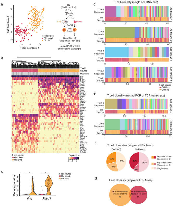
The mammalian brain contains neurogenic niches that comprise neural stem cells and other cell types. Neurogenic niches become less functional with age, but how they change during ageing remains unclear. Here we perform single-cell RNA sequencing of young and old neurogenic niches in mice. The analysis of 14,685 single-cell transcriptomes reveals a decrease in activated neural stem cells, changes in endothelial cells and microglia, and an infiltration of T cells in old neurogenic niches. T cells in old brains are clonally expanded and are generally distinct from those in old blood, which suggests that they may experience specific antigens. T cells in old brains also express interferon-γ, and the subset of neural stem cells that has a high interferon response shows decreased proliferation in vivo. We find that T cells can inhibit the proliferation of neural stem cells in co-cultures and in vivo, in part by secreting interferon-γ. Our study reveals an interaction between T cells and neural stem cells in old brains, opening potential avenues through which to counteract age-related decline in brain function.

摘要

哺乳动物大脑包含神经发生龛,其中包括神经干细胞和其他细胞类型。神经发生龛随着年龄的增长而功能下降,但它们在衰老过程中如何变化尚不清楚。在这里,我们对年轻和年老的小鼠神经发生龛进行了单细胞 RNA 测序。对 14685 个单细胞转录组的分析揭示了激活的神经干细胞减少、内皮细胞和小胶质细胞变化以及 T 细胞在老年神经发生龛中的浸润。老年大脑中的 T 细胞克隆扩增,通常与老年血液中的 T 细胞不同,这表明它们可能经历特定的抗原。老年大脑中的 T 细胞也表达干扰素-γ,具有高干扰素反应的神经干细胞亚群在体内的增殖减少。我们发现 T 细胞可以在共培养物和体内抑制神经干细胞的增殖,部分是通过分泌干扰素-γ。我们的研究揭示了老年大脑中 T 细胞和神经干细胞之间的相互作用,为对抗与年龄相关的大脑功能下降开辟了潜在途径。

https://cdn.ncbi.nlm.nih.gov/pmc/blobs/8994/7111535/ddc7b1b5584d/nihms-1531024-f0004.jpg
https://cdn.ncbi.nlm.nih.gov/pmc/blobs/8994/7111535/54533cf58c8c/nihms-1531024-f0005.jpg
https://cdn.ncbi.nlm.nih.gov/pmc/blobs/8994/7111535/870193a9da1a/nihms-1531024-f0006.jpg
https://cdn.ncbi.nlm.nih.gov/pmc/blobs/8994/7111535/8df56e0df7a2/nihms-1531024-f0007.jpg
https://cdn.ncbi.nlm.nih.gov/pmc/blobs/8994/7111535/a5c2175d647f/nihms-1531024-f0008.jpg
https://cdn.ncbi.nlm.nih.gov/pmc/blobs/8994/7111535/91b6b7b17cf1/nihms-1531024-f0009.jpg
https://cdn.ncbi.nlm.nih.gov/pmc/blobs/8994/7111535/654bbdaf9fe5/nihms-1531024-f0010.jpg
https://cdn.ncbi.nlm.nih.gov/pmc/blobs/8994/7111535/9241fa56bb60/nihms-1531024-f0011.jpg
https://cdn.ncbi.nlm.nih.gov/pmc/blobs/8994/7111535/fbaccd1dac7e/nihms-1531024-f0001.jpg
https://cdn.ncbi.nlm.nih.gov/pmc/blobs/8994/7111535/6c20be108cd7/nihms-1531024-f0002.jpg
https://cdn.ncbi.nlm.nih.gov/pmc/blobs/8994/7111535/a138331937e5/nihms-1531024-f0003.jpg
https://cdn.ncbi.nlm.nih.gov/pmc/blobs/8994/7111535/ddc7b1b5584d/nihms-1531024-f0004.jpg

相似文献

1
Single-cell analysis reveals T cell infiltration in old neurogenic niches.单细胞分析揭示了 T 细胞在衰老神经发生龛中的浸润。
Nature. 2019 Jul;571(7764):205-210. doi: 10.1038/s41586-019-1362-5. Epub 2019 Jul 3.
2
Vascular-derived TGF-β increases in the stem cell niche and perturbs neurogenesis during aging and following irradiation in the adult mouse brain.血管源性 TGF-β在干细胞龛中增加,并在成年小鼠大脑衰老和辐照后扰乱神经发生。
EMBO Mol Med. 2013 Apr;5(4):548-62. doi: 10.1002/emmm.201202197. Epub 2013 Mar 25.
3
Akhirin regulates the proliferation and differentiation of neural stem cells/progenitor cells at neurogenic niches in mouse brain.Akhirin 在小鼠脑的神经发生龛中调节神经干细胞/祖细胞的增殖和分化。
Dev Growth Differ. 2020 Feb;62(2):97-107. doi: 10.1111/dgd.12646. Epub 2020 Jan 13.
4
Neurogenic Niche Microglia Undergo Positional Remodeling and Progressive Activation Contributing to Age-Associated Reductions in Neurogenesis.神经源性微环境小胶质细胞经历位置重塑和渐进性激活,导致与年龄相关的神经发生减少。
Stem Cells Dev. 2016 Apr 1;25(7):542-55. doi: 10.1089/scd.2015.0319. Epub 2016 Mar 16.
5
NTPDase2 and purinergic signaling control progenitor cell proliferation in neurogenic niches of the adult mouse brain.NTPDase2和嘌呤能信号传导控制成年小鼠脑海马神经干细胞龛中的祖细胞增殖。
Stem Cells. 2015 Jan;33(1):253-64. doi: 10.1002/stem.1846.
6
Single-Cell Transcriptomics Characterizes Cell Types in the Subventricular Zone and Uncovers Molecular Defects Impairing Adult Neurogenesis.单细胞转录组学描绘了脑室下区的细胞类型,并揭示了影响成年神经发生的分子缺陷。
Cell Rep. 2018 Nov 27;25(9):2457-2469.e8. doi: 10.1016/j.celrep.2018.11.003.
7
Neural transmembrane protease and endothelial Gs protein activation in cell contact-dependent signaling between neural stem/progenitor cells and brain endothelial cells.神经跨膜蛋白酶和内皮 Gs 蛋白在神经干细胞/祖细胞与脑内皮细胞之间的细胞接触依赖性信号转导中的激活作用。
J Biol Chem. 2012 Jun 29;287(27):22497-508. doi: 10.1074/jbc.M111.330589. Epub 2012 May 10.
8
Home sweet home: the neural stem cell niche throughout development and after injury.温馨的家:神经干细胞在发育过程中和损伤后的龛位。
Cell Tissue Res. 2018 Jan;371(1):125-141. doi: 10.1007/s00441-017-2658-0. Epub 2017 Aug 3.
9
Physiological Interactions between Microglia and Neural Stem Cells in the Adult Subependymal Niche.成年室管膜下区小胶质细胞与神经干细胞的生理相互作用。
Neuroscience. 2019 May 1;405:77-91. doi: 10.1016/j.neuroscience.2019.01.009. Epub 2019 Jan 21.
10
The Role of the Microenvironmental Niche in Declining Stem-Cell Functions Associated with Biological Aging.微环境生态位在与生物衰老相关的干细胞功能衰退中的作用
Cold Spring Harb Perspect Med. 2015 Dec 1;5(12):a025874. doi: 10.1101/cshperspect.a025874.

引用本文的文献

1
Haematopoietic ageing in health and lifespan.健康与寿命中的造血衰老
Nat Cell Biol. 2025 Aug 25. doi: 10.1038/s41556-025-01739-1.
2
The Inflammatory Bridge Between Type 2 Diabetes and Neurodegeneration: A Molecular Perspective.2型糖尿病与神经退行性变之间的炎症桥梁:分子视角
Int J Mol Sci. 2025 Aug 5;26(15):7566. doi: 10.3390/ijms26157566.
3
Metabolic landscape uncovers remodeling of T cell immunity affected by fatty acid desaturase in Parkinson's disease at single-cell resolution.代谢图谱在单细胞分辨率下揭示了帕金森病中受脂肪酸去饱和酶影响的T细胞免疫重塑。

本文引用的文献

1
Single-cell transcriptomics reveals gene signatures and alterations associated with aging in distinct neural stem/progenitor cell subpopulations.单细胞转录组学揭示了与不同神经干细胞/祖细胞亚群衰老相关的基因特征和改变。
Protein Cell. 2018 Apr;9(4):351-364. doi: 10.1007/s13238-017-0450-2. Epub 2017 Jul 26.
2
Isolation of CNS-infiltrating and Resident Microglial Cells.中枢神经系统浸润性和驻留小胶质细胞的分离。
Bio Protoc. 2015 Jan 20;5(2). doi: 10.21769/bioprotoc.1385.
3
IFN gamma regulates proliferation and neuronal differentiation by STAT1 in adult SVZ niche.
BMC Biol. 2025 Aug 6;23(1):245. doi: 10.1186/s12915-025-02358-w.
4
Human brain vascular multi-omics elucidates disease-risk associations.人类脑血管多组学揭示疾病风险关联。
Neuron. 2025 Jul 23. doi: 10.1016/j.neuron.2025.07.001.
5
Unraveling regulatory feedback mechanisms in adult neurogenesis through mathematical modelling.通过数学建模揭示成体神经发生中的调控反馈机制。
NPJ Syst Biol Appl. 2025 Jul 26;11(1):82. doi: 10.1038/s41540-025-00563-5.
6
How stem cells respond to infection, inflammation and ageing.干细胞如何应对感染、炎症和衰老。
Nat Rev Immunol. 2025 Jul 24. doi: 10.1038/s41577-025-01203-z.
7
CHI3L1/YKL-40 signaling inhibits neurogenesis in models of Alzheimer's disease.几丁质酶3样蛋白1/壳多糖酶-3样蛋白40(CHI3L1/YKL-40)信号通路在阿尔茨海默病模型中抑制神经发生。
Sci Adv. 2025 Jul 18;11(29):eadv1492. doi: 10.1126/sciadv.adv1492.
8
Cellular senescence, neuroinflammation, and microRNAs: Possible interactions driving aging and neurodegeneration in the hippocampal neurogenic niche.细胞衰老、神经炎症与微小RNA:驱动海马神经发生微环境衰老和神经退行性变的可能相互作用
Aging Brain. 2025 Jun 12;8:100141. doi: 10.1016/j.nbas.2025.100141. eCollection 2025.
9
Comprehensive immune ageing reveals TREM2/TIM3 myeloid cells drive brain immune evasion.全面的免疫衰老揭示TREM2/TIM3髓样细胞驱动脑部免疫逃逸。
EBioMedicine. 2025 Jun 27;118:105833. doi: 10.1016/j.ebiom.2025.105833.
10
Hallmarks of stem cell aging.干细胞衰老的特征。
Cell Stem Cell. 2025 Jul 3;32(7):1038-1054. doi: 10.1016/j.stem.2025.06.004. Epub 2025 Jun 24.
干扰素γ通过信号转导和转录激活因子1(STAT1)调节成年侧脑室下区生态位中的细胞增殖和神经元分化。
Front Cell Neurosci. 2015 Jul 13;9:270. doi: 10.3389/fncel.2015.00270. eCollection 2015.