Hu Yan, Guo Tie-Cheng, Zhang Xiang-Yu, Tian Jun, Lu Yin-Shan
Department of Rehabilitation Medicine, Tongji Hospital, Tongji Medical College, Huazhong University of Science & Technology; Department of Rehabilitation Medicine, Zhongnan Hospital of Wuhan University, Wuhan, Hubei Province, China.
Department of Rehabilitation Medicine, Tongji Hospital, Tongji Medical College, Huazhong University of Science & Technology, Wuhan, Hubei Province, China.
Neural Regen Res. 2019 Nov;14(11):1968-1976. doi: 10.4103/1673-5374.259618.
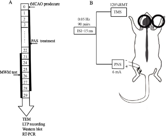
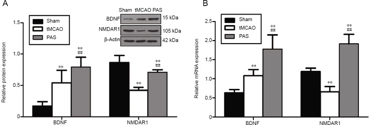
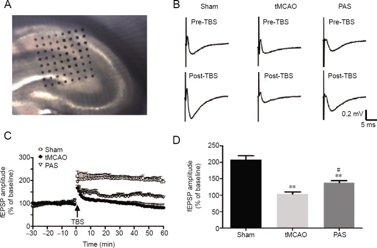
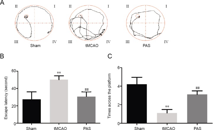
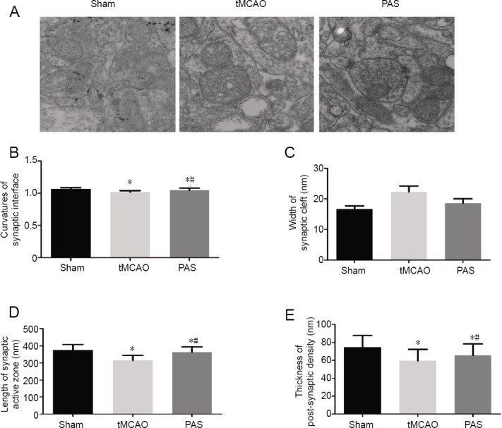
Paired associative stimulation is a relatively new non-invasive brain stimulation technique that combines transcranial magnetic stimulation and peripheral nerve stimulation. The effects of paired associative stimulation on the excitability of the cerebral cortex can vary according to the time interval between the transcranial magnetic stimulation and peripheral nerve stimulation. We established a model of cerebral ischemia in rats via transient middle cerebral artery occlusion. We administered paired associative stimulation with a frequency of 0.05 Hz 90 times over 4 weeks. We then evaluated spatial learning and memory using the Morris water maze. Changes in the cerebral ultra-structure and synaptic plasticity were assessed via transmission electron microscopy and a 64-channel multi-electrode array. We measured mRNA and protein expression levels of brain-derived neurotrophic factor and N-methyl-D-aspartate receptor 1 in the hippocampus using a real-time polymerase chain reaction and western blot assay. Paired associative stimulation treatment significantly improved learning and memory in rats subjected to cerebral ischemia. The ultra-structures of synapses in the CA1 area of the hippocampus in rats subjected to cerebral ischemia were restored by paired associative stimulation. Long-term potentiation at synapses in the CA3 and CA1 regions of the hippocampus was enhanced as well. The protein and mRNA expression of brain-derived neurotrophic factor and N-methyl-D-aspartate receptor 1 increased after paired associative stimulation treatment. These data indicate that paired associative stimulation can protect cognition after cerebral ischemia. The observed effect may be mediated by increases in the mRNA and protein expression of brain-derived neurotrophic factor and N-methyl-D-aspartate receptor 1, and by enhanced synaptic plasticity in the CA1 area of the hippocampus. The animal experiments were approved by the Animal Ethics Committee of Tongji Medical College, Huazhong University of Science & Technology, China (approval No. TJ-A20151102) on July 11, 2015.
配对联想刺激是一种相对较新的非侵入性脑刺激技术,它结合了经颅磁刺激和外周神经刺激。配对联想刺激对大脑皮层兴奋性的影响会因经颅磁刺激和外周神经刺激之间的时间间隔而有所不同。我们通过短暂性大脑中动脉闭塞建立了大鼠脑缺血模型。我们在4周内以0.05Hz的频率进行了90次配对联想刺激。然后使用莫里斯水迷宫评估空间学习和记忆。通过透射电子显微镜和64通道多电极阵列评估脑超微结构和突触可塑性的变化。我们使用实时聚合酶链反应和蛋白质印迹法测量海马中脑源性神经营养因子和N-甲基-D-天冬氨酸受体1的mRNA和蛋白质表达水平。配对联想刺激治疗显著改善了脑缺血大鼠的学习和记忆。配对联想刺激使脑缺血大鼠海马CA1区突触的超微结构得以恢复。海马CA3和CA1区突触的长时程增强也得到了增强。配对联想刺激治疗后,脑源性神经营养因子和N-甲基-D-天冬氨酸受体1的蛋白质和mRNA表达增加。这些数据表明,配对联想刺激可以保护脑缺血后的认知。观察到的效果可能是由脑源性神经营养因子和N-甲基-D-天冬氨酸受体1的mRNA和蛋白质表达增加以及海马CA1区突触可塑性增强介导的。动物实验于2015年7月11日获得中国华中科技大学同济医学院动物伦理委员会批准(批准号:TJ-A20151102)。