Numata Tomohiro, Sato-Numata Kaori, Okada Yasunobu
Department of Physiology, Graduate School of Medical Sciences, Fukuoka University, Fukuoka, Japan.
Japan Society for the Promotion of Science, Tokyo, Japan.
Physiol Rep. 2019 Jul;7(13):e14157. doi: 10.14814/phy2.14157.
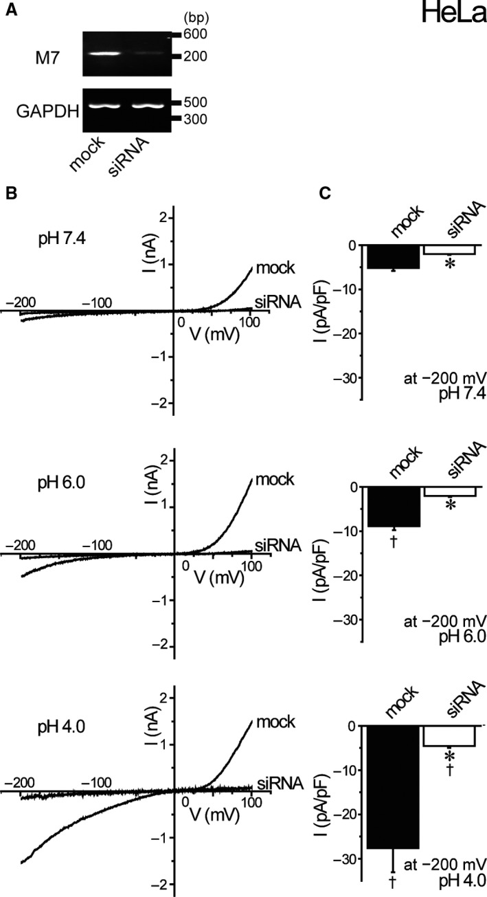
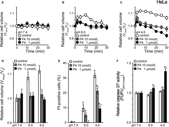
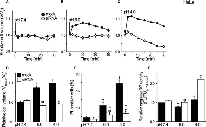
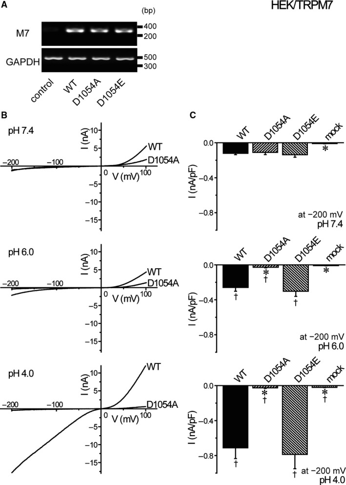
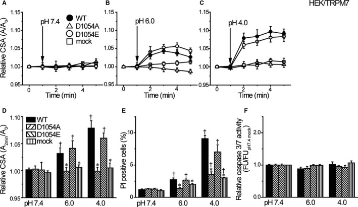
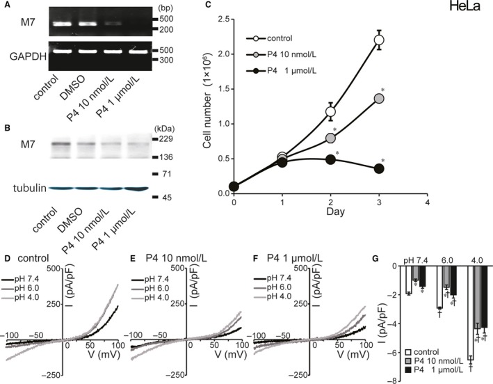
Because intravaginal pH is strongly acidic, it is important to investigate the effects of acidosis on cervical cancer cells. Recently, in response to strong acidosis, human cervical cancer HeLa cells were shown to exhibit necrosis after showing persistent cell swelling induced by Cl influx. Since cation influx should be accompanied with Cl influx to drive water inflow causing cell swelling, we here studied on the nature of acidotoxic cation conductance. The mRNA/protein expression was assessed by RT-PCR and Western blotting. Ionic currents were measured by patch-clamping techniques. Cell counting/viability and colorimetric assays were applied to assess proliferation rate and caspase 3/7 activity, respectively. Cell volume and size were measured by electronic sizing and video-microscopic measurements, respectively. Acid exposure enhanced TRPM7 activity endogenously expressed in HeLa cells and exogenously overexpressed in HEK293T cells. Gene silencing of TRPM7 abolished acid-induced cell swelling and necrosis but rather induced activation of apoptotic caspase 3/7 in HeLa cells. Overexpression with the pore charge-neutralizing D1054A mutant suppressed acid-enhanced cation currents, acid-induced cell swelling, and acidotoxic necrosis in HEK293T cells. Progesterone treatment was surprisingly found to suppress molecular and functional expression of TRPM7 and cell proliferation in HeLa cells. Furthermore, in the progesterone-treated cells, acid exposure did not induce persistent cell swelling followed by necrosis but induced persistent cell shrinkage and apoptotic cell death. These results indicate that in the human cervical cancer cells, TRPM7 is essentially involved in acidotoxic necrotic cell death, and progesterone inhibits TRPM7 expression thereby inhibiting acidotoxic necrosis by switching to apoptosis.
由于阴道内pH值呈强酸性,因此研究酸中毒对宫颈癌细胞的影响很重要。最近,研究发现,在受到强酸中毒刺激后,人宫颈癌HeLa细胞先是因Cl⁻内流诱导持续细胞肿胀,随后出现坏死。由于阳离子内流应伴随Cl⁻内流以驱动水流导致细胞肿胀,因此我们在此研究了酸毒性阳离子电导的性质。通过RT-PCR和蛋白质印迹法评估mRNA/蛋白质表达。采用膜片钳技术测量离子电流。分别应用细胞计数/活力检测和比色法评估增殖率和半胱天冬酶3/7活性。分别通过电子大小测量和视频显微镜测量来测定细胞体积和大小。酸暴露增强了HeLa细胞内源性表达以及HEK293T细胞外源性过表达的TRPM7活性。TRPM7基因沉默消除了酸诱导的细胞肿胀和坏死,但反而诱导了HeLa细胞中凋亡性半胱天冬酶3/7的激活。用孔电荷中和的D1054A突变体过表达可抑制HEK293T细胞中酸增强的阳离子电流、酸诱导的细胞肿胀和酸毒性坏死。令人惊讶的是,发现孕酮处理可抑制HeLa细胞中TRPM7的分子和功能表达以及细胞增殖。此外,在孕酮处理的细胞中,酸暴露不会诱导持续的细胞肿胀继而坏死,而是诱导持续的细胞收缩和凋亡性细胞死亡。这些结果表明,在人宫颈癌细胞中,TRPM7本质上参与酸毒性坏死性细胞死亡,而孕酮抑制TRPM7表达,从而通过转变为凋亡来抑制酸毒性坏死。