• 文献检索
  • 文档翻译
  • 深度研究
  • 学术资讯
  • Suppr Zotero 插件Zotero 插件
  • 邀请有礼
  • 套餐&价格
  • 历史记录
应用&插件
Suppr Zotero 插件Zotero 插件浏览器插件Mac 客户端Windows 客户端微信小程序
定价
高级版会员购买积分包购买API积分包
服务
文献检索文档翻译深度研究API 文档MCP 服务
关于我们
关于 Suppr公司介绍联系我们用户协议隐私条款
关注我们

Suppr 超能文献

核心技术专利:CN118964589B侵权必究
粤ICP备2023148730 号-1Suppr @ 2026

文献检索

告别复杂PubMed语法,用中文像聊天一样搜索,搜遍4000万医学文献。AI智能推荐,让科研检索更轻松。

立即免费搜索

文件翻译

保留排版,准确专业,支持PDF/Word/PPT等文件格式,支持 12+语言互译。

免费翻译文档

深度研究

AI帮你快速写综述,25分钟生成高质量综述,智能提取关键信息,辅助科研写作。

立即免费体验

大鼠牙囊干细胞不同传代培养时的超微结构形态学特征

Ultrastructure Morphological Characterization of Different Passages of Rat Dental Follicle Stem Cells at Culture.

作者信息

Al-Bagdadi Fakhri A, Barona Humberto M, Martinez-Ceballos Eduardo, Yao Shaomian

机构信息

Department of Comparative Biomedical Sciences, School of Veterinary Medicine, Louisiana State University, Baton Rouge, LA, USA.

Department of Mathematics, Southern University and A and M College, Baton Rouge, LA, USA.

出版信息

J Microsc Ultrastruct. 2019 Apr-Jun;7(2):57-64. doi: 10.4103/JMAU.JMAU_44_18.

DOI:10.4103/JMAU.JMAU_44_18
PMID:31293886
原文链接:https://pmc.ncbi.nlm.nih.gov/articles/PMC6585478/
Abstract

INTRODUCTION

Stem cells play important roles in tissue renewal and repair. Tissue-derived stem cells have been demonstrated for their applications in tissue engineering and regenerative medicine. Expansion of primary stem cells isolated from tissues to a large quantity through culture is needed for application of the stem cells. However, it is known that tissue stem cells commonly reduce or lose their stemness properties during in vitro culture. In this study, we assessed ultrastructural changes of rat dental follicle stem cells (DFSCs) during in vitro culture. It is our attempt to explain the loss of stemness properties in cultured tissue-stem cells at the ultrastructural level.

METHOD

DFSCs was isolated from first molars of Sprague Dawley rat pups and cultured in medium consisting of alpha-MEM plus 20% FBS. Cells were passaged at 1 to 3 ratio at 90% confluence, and collected at passages 3, 6, 7 and 9 for assessment of ultrastructure morphology by transmission electron microscopy.

RESULTS

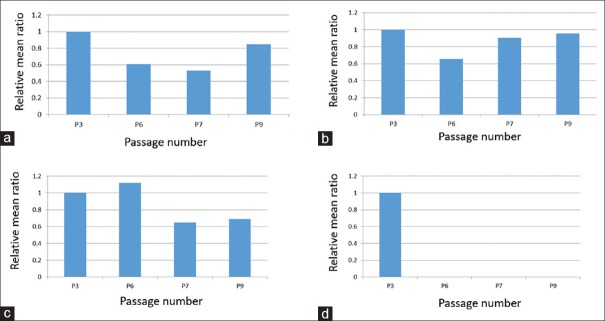
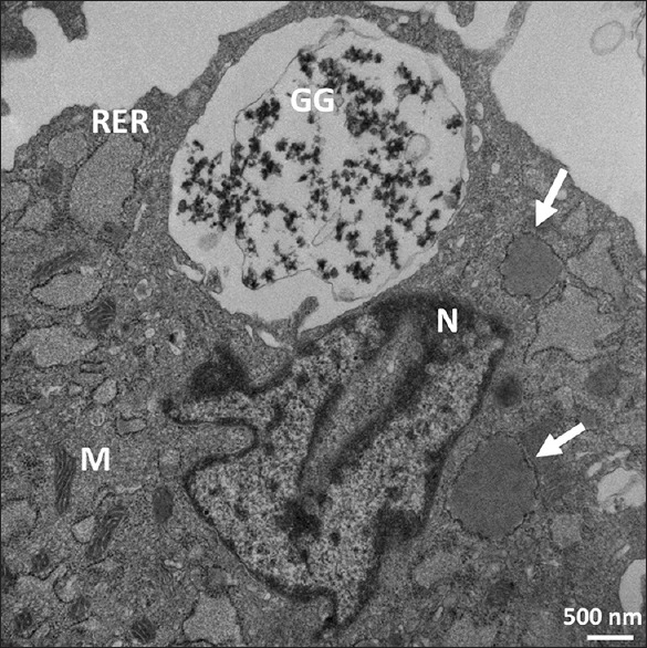
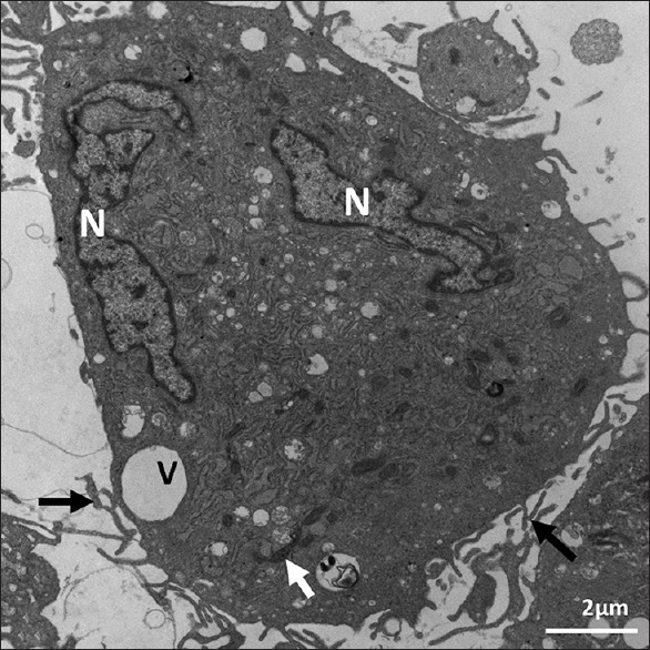
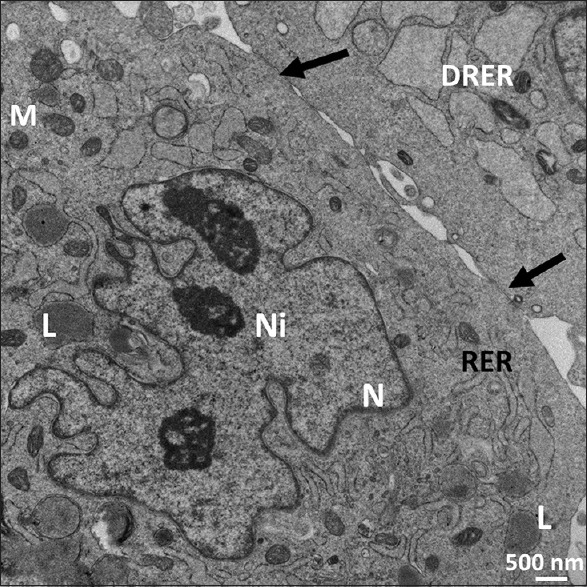
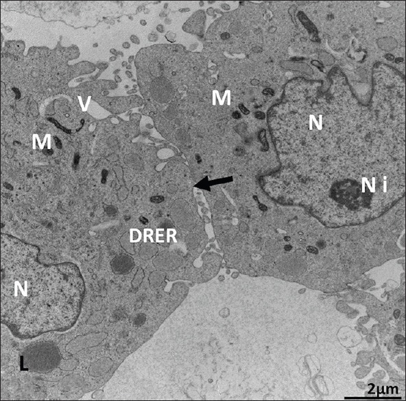
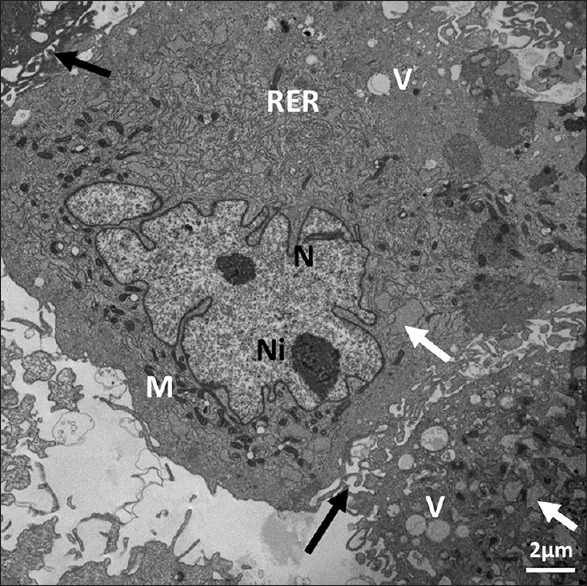
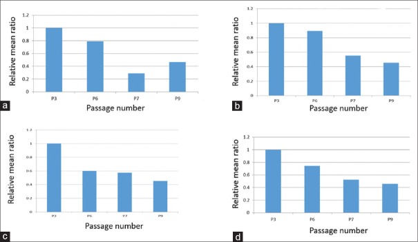
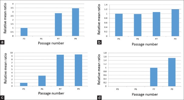
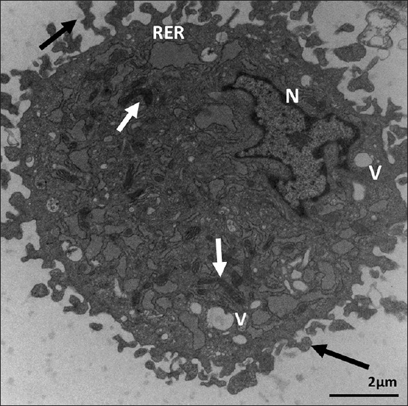
Of the four passages (3, 6, 7, and 9) examined, dilated rough endoplasmic reticulum (RER) was abundant in Passage 3 but less so in Passages 6, 7, and 9. The dilated RER contained lipid in Passages 3, 7, and 9. The mono- and polyribosomes in Passages 3 and 6 were located between the mitochondria and the RER. Mono- and polyribosomes were abundant in Passage 7, although mainly monoribosomes were present in Passage 9. Membrane-bound glycogen granules were in vacuoles bulging off the cells in Passage 3. Some glycogen granules were grouped in the periphery of a stem cell in Passage 9. Nuclei shapes were irregular and mainly euchromatic in Passages 6, 7, and 9. The mitochondria were dark and scarce in Passage 9; irregular, small, and dark in Passage 7; and small and rounded in Passage 6, and they were spread in the cytoplasm away from the nucleus in Passage 3. Cell contacts were seen in Passages 6, 7, and 9. The ultrastructure morphology of the examined DFScs was not very different from the morphology criteria of the undifferentiated cells. Large vacuoles in Passage 3 were mainly at the periphery of the cell, with the small vacuoles in the cell center. Small vacuoles were scattered in the cell center of Passage 6 and the larger ones were observed at the cell's periphery.

CONCLUSIONS

We observed the following ultrastructural changes: decreases of fine cell cytoplasmic processes, dilated cytoplasmic vacuoles, cytoplasmic pinocytotic vesicles, and nuclear heterochromatin with increasing cell passage number. Conversely, mean ratios of lipid globules, nuclear euchromatin, irregular nuclear shape, and cell contact between cells were increased with passage number. The observations may suggest an increase in committed cells among the population after long-term culture of DFSCs.

https://cdn.ncbi.nlm.nih.gov/pmc/blobs/2609/6585478/2b31a9db0ff4/JMAU-7-57-g011.jpg
https://cdn.ncbi.nlm.nih.gov/pmc/blobs/2609/6585478/a4454e552686/JMAU-7-57-g003.jpg
https://cdn.ncbi.nlm.nih.gov/pmc/blobs/2609/6585478/045b37214ddd/JMAU-7-57-g004.jpg
https://cdn.ncbi.nlm.nih.gov/pmc/blobs/2609/6585478/aecf079b4ef2/JMAU-7-57-g005.jpg
https://cdn.ncbi.nlm.nih.gov/pmc/blobs/2609/6585478/47adb3815ea2/JMAU-7-57-g006.jpg
https://cdn.ncbi.nlm.nih.gov/pmc/blobs/2609/6585478/a6cbb2b6c0cb/JMAU-7-57-g007.jpg
https://cdn.ncbi.nlm.nih.gov/pmc/blobs/2609/6585478/2660e45992cd/JMAU-7-57-g008.jpg
https://cdn.ncbi.nlm.nih.gov/pmc/blobs/2609/6585478/6bf42c433fe6/JMAU-7-57-g009.jpg
https://cdn.ncbi.nlm.nih.gov/pmc/blobs/2609/6585478/011d3f8bc384/JMAU-7-57-g010.jpg
https://cdn.ncbi.nlm.nih.gov/pmc/blobs/2609/6585478/2b31a9db0ff4/JMAU-7-57-g011.jpg
摘要

引言

干细胞在组织更新和修复中发挥着重要作用。组织来源的干细胞已在组织工程和再生医学中得到应用。为了应用干细胞,需要通过培养将从组织中分离出的原代干细胞大量扩增。然而,已知组织干细胞在体外培养过程中通常会降低或丧失其干性特性。在本研究中,我们评估了大鼠牙囊干细胞(DFSCs)在体外培养过程中的超微结构变化。我们试图在超微结构水平上解释培养的组织干细胞干性特性的丧失。

方法

从Sprague Dawley大鼠幼崽的第一磨牙中分离DFSCs,并在含有α-MEM加20%胎牛血清的培养基中培养。细胞在90%汇合时以1:3的比例传代,并在第3、6、7和9代收集,通过透射电子显微镜评估超微结构形态。

结果

在所检查的四代(第3、6、7和9代)中,第3代粗面内质网(RER)扩张丰富,而第6、7和9代则较少。第3、7和9代扩张的RER中含有脂质。第3和6代的单核糖体和多核糖体位于线粒体和RER之间。第7代单核糖体和多核糖体丰富,尽管第9代主要是单核糖体。第3代膜结合的糖原颗粒存在于从细胞突出的液泡中。第9代一些糖原颗粒聚集在干细胞周边。第6、7和9代细胞核形状不规则且主要为常染色质。第9代线粒体暗且稀少;第7代不规则、小且暗;第6代小且呈圆形,它们在第3代中分散在远离细胞核的细胞质中。第6、7和9代可见细胞间接触。所检查的DFSCs的超微结构形态与未分化细胞的形态标准差异不大。第3代大液泡主要位于细胞周边,小液泡位于细胞中心。第6代小液泡散在细胞中心,较大的液泡在细胞周边观察到。

结论

我们观察到以下超微结构变化:随着细胞传代次数增加,细胞精细的细胞质突起、扩张的细胞质液泡、细胞质吞饮小泡和核异染色质减少。相反,脂质球、核常染色质、不规则核形状以及细胞间接触的平均比例随传代次数增加。这些观察结果可能表明DFSCs长期培养后群体中定向分化细胞增加。

相似文献

1
Ultrastructure Morphological Characterization of Different Passages of Rat Dental Follicle Stem Cells at Culture.大鼠牙囊干细胞不同传代培养时的超微结构形态学特征
J Microsc Ultrastruct. 2019 Apr-Jun;7(2):57-64. doi: 10.4103/JMAU.JMAU_44_18.
2
Ultrastructural comparison of porcine putative embryonic stem cells derived by in vitro fertilization and somatic cell nuclear transfer.体外受精和体细胞核移植获得的猪假定胚胎干细胞的超微结构比较
J Reprod Dev. 2016 Apr 22;62(2):177-85. doi: 10.1262/jrd.2015-124. Epub 2016 Jan 28.
3
Morphology of in vitro expanded human muscle-derived stem cells.体外扩增的人肌肉衍生干细胞的形态学
Biomed Pap Med Fac Univ Palacky Olomouc Czech Repub. 2008 Dec;152(2):235-8. doi: 10.5507/bp.2008.036.
4
[ULTRASTRUCTURAL CHANGES OF THE STEM CELLS IN THE CYCLE MONOLAYER--SPHERES--MONOLAYER].
Tsitologiia. 2016;58(1):16-22.
5
Defensive ink pigment processing and secretion in Aplysia californica: concentration and storage of phycoerythrobilin in the ink gland.加州海兔的防御性墨汁色素加工与分泌:藻红胆素在墨腺中的浓缩与储存
J Exp Biol. 1998 May;201(Pt 10):1595-613. doi: 10.1242/jeb.201.10.1595.
6
Expression of bone morphogenetic protein-6 in dental follicle stem cells and its effect on osteogenic differentiation.骨形态发生蛋白-6在牙囊干细胞中的表达及其对成骨分化的影响。
Cells Tissues Organs. 2013;198(6):438-47. doi: 10.1159/000360275. Epub 2014 Apr 8.
7
Small extracellular vesicles from dental follicle stem cells provide biochemical cues for periodontal tissue regeneration.牙囊干细胞来源的小细胞外囊泡为牙周组织再生提供生化线索。
Stem Cell Res Ther. 2022 Mar 3;13(1):92. doi: 10.1186/s13287-022-02767-6.
8
[Establishment of cultivating technology of dental follicle cells of rat and their identifying].[大鼠牙囊细胞培养技术的建立及其鉴定]
Hua Xi Kou Qiang Yi Xue Za Zhi. 2004 Feb;22(1):19-22.
9
Ultrastructure of cultured cells from Schistosoma japonicum.
Acta Trop. 2002 May;82(2):225-34. doi: 10.1016/s0001-706x(02)00014-1.
10
Hemocytes ultrastructure of Aedes aegypti (Diptera: Culicidae).埃及伊蚊(双翅目:蚊科)血细胞的超微结构
Micron. 2008;39(2):184-9. doi: 10.1016/j.micron.2007.01.003. Epub 2007 Jan 21.

引用本文的文献

1
Optimal good manufacturing practice-compliant production of dental follicle stem cell sheet and its application in Sprague-Dawley rat periodontitis.符合药品生产质量管理规范的牙囊干细胞片的优化生产及其在斯普拉格-道利大鼠牙周炎中的应用
World J Stem Cells. 2025 May 26;17(5):104116. doi: 10.4252/wjsc.v17.i5.104116.
2
Characterizing Organelles in Live Stem Cells Using Label-Free Optical Diffraction Tomography.使用无标记光衍射层析术对活干细胞中的细胞器进行特征描述。
Mol Cells. 2021 Nov 30;44(11):851-860. doi: 10.14348/molcells.2021.0190.

本文引用的文献

1
Ultrastructural visualization of the Mesenchymal-to-Epithelial Transition during reprogramming of human fibroblasts to induced pluripotent stem cells.人类成纤维细胞重编程为诱导多能干细胞过程中上皮-间充质转化的超微结构可视化
Stem Cell Res. 2015 Jan;14(1):39-53. doi: 10.1016/j.scr.2014.11.003. Epub 2014 Nov 29.
2
Multipotent to pluripotent properties of adult stem cells.成体干细胞的多能性向全能性特性
Stem Cells Int. 2013;2013:813780. doi: 10.1155/2013/813780. Epub 2013 Dec 15.
3
An efficient and reproducible process for transmission electron microscopy (TEM) of rare cell populations.
一种高效且可重现的用于透射电子显微镜(TEM)稀有细胞群体成像的方法。
J Immunol Methods. 2014 Feb;404:87-90. doi: 10.1016/j.jim.2013.11.025. Epub 2013 Dec 1.
4
Engineering stem cell niches in bioreactors.在生物反应器中构建干细胞生态位。
World J Stem Cells. 2013 Oct 26;5(4):124-35. doi: 10.4252/wjsc.v5.i4.124.
5
Microenvironment at tissue injury, a key focus for efficient stem cell therapy: A discussion of mesenchymal stem cells.组织损伤微环境:高效干细胞治疗的关键关注点:间充质干细胞的讨论。
World J Stem Cells. 2009 Dec 31;1(1):3-7. doi: 10.4252/wjsc.v1.i1.3.
6
Therapeutic potential of appropriately evaluated safe-induced pluripotent stem cells for spinal cord injury.安全评估诱导多能干细胞治疗脊髓损伤的潜力。
Proc Natl Acad Sci U S A. 2010 Jul 13;107(28):12704-9. doi: 10.1073/pnas.0910106107. Epub 2010 Jul 6.
7
Differentiation potential of stem cells from human dental origin - promise for tissue engineering.人源性牙齿干细胞的分化潜能——组织工程学的应用前景。
J Physiol Pharmacol. 2009 Dec;60 Suppl 7:167-75.
8
Up-scaling single cell-inoculated suspension culture of human embryonic stem cells.扩大人胚胎干细胞单细胞接种悬浮培养规模。
Stem Cell Res. 2010 May;4(3):165-79. doi: 10.1016/j.scr.2010.03.001. Epub 2010 Mar 12.
9
Irradiation of adult human dental pulp stem cells provokes activation of p53, cell cycle arrest, and senescence but not apoptosis.辐照成人牙髓干细胞可引发 p53 激活、细胞周期停滞和衰老,但不会引发细胞凋亡。
Stem Cells Dev. 2010 Dec;19(12):1855-62. doi: 10.1089/scd.2009.0449. Epub 2010 Sep 13.
10
Characterization of mesenchymal stem cells from rat bone marrow: ultrastructural properties, differentiation potential and immunophenotypic markers.大鼠骨髓间充质干细胞的鉴定:超微结构特性、分化潜能和免疫表型标志物。
Histochem Cell Biol. 2009 Nov;132(5):533-46. doi: 10.1007/s00418-009-0629-6. Epub 2009 Aug 14.