Cárdenas-Rivera Alfredo, Campero-Romero Aura N, Heras-Romero Yessica, Penagos-Puig Andrés, Rincón-Heredia Ruth, Tovar-Y-Romo Luis B
Division of Neuroscience, Instituto de Fisiología Celular, Universidad Nacional Autónoma de México, Mexico City, Mexico.
Microscopy Core Unit, Instituto de Fisiología Celular, Universidad Nacional Autónoma de México, Mexico City, Mexico.
Front Cell Neurosci. 2019 Jul 2;13:270. doi: 10.3389/fncel.2019.00270. eCollection 2019.
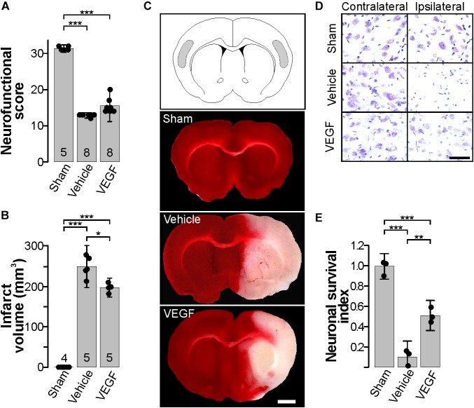
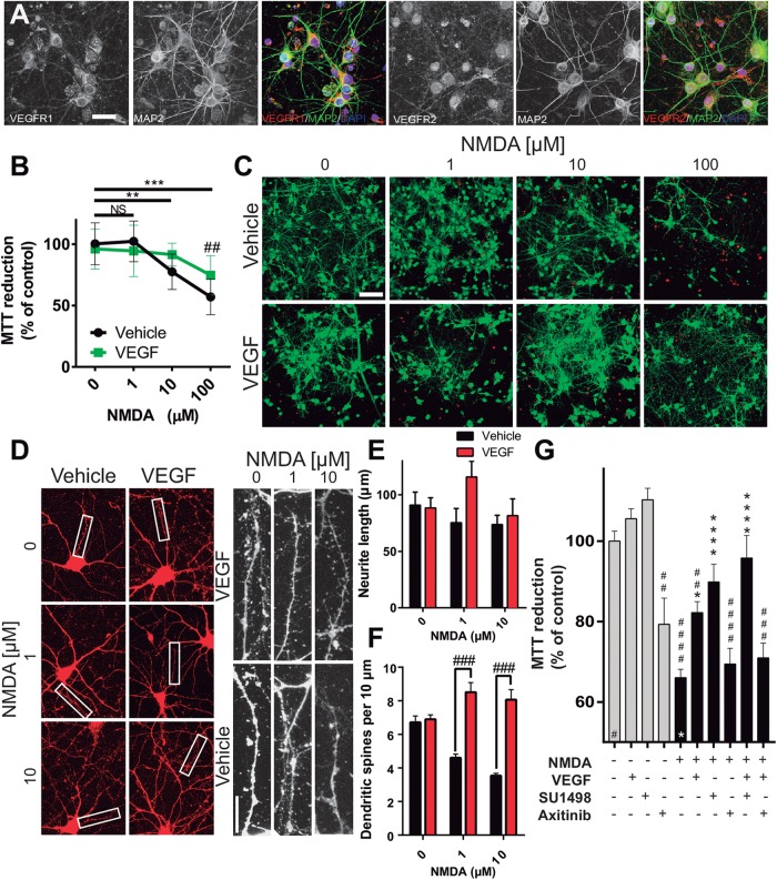
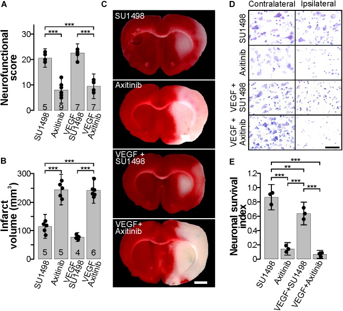
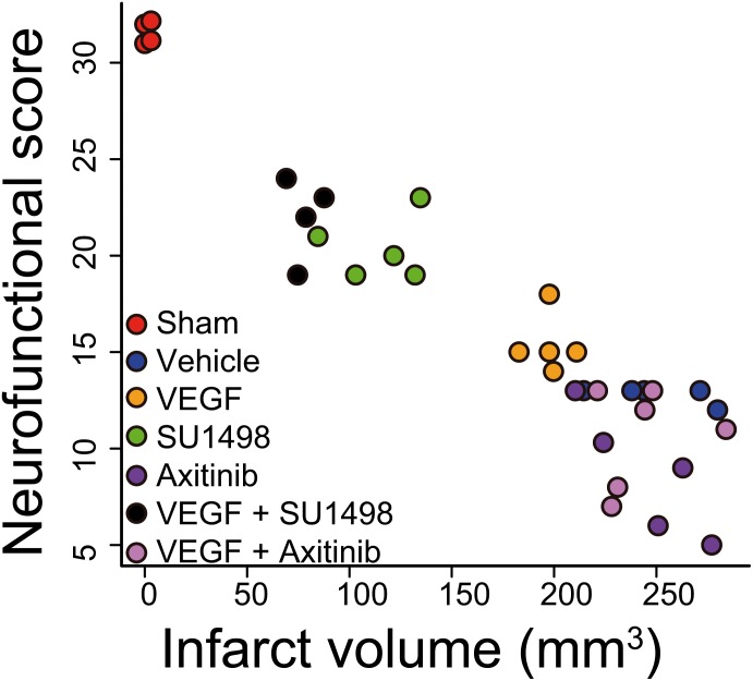
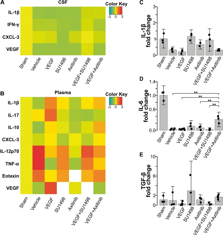
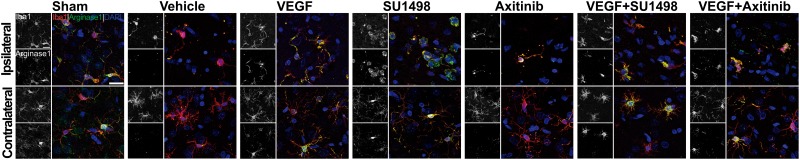
Vascular endothelial growth factor (VEGF) has long been connected to the development of tissue lesion following ischemic stroke. Contradictory findings either situate VEGF as a promoter of large infarct volumes or as a potential attenuator of damage due to its well documented neuroprotective capability. The core of this discrepancy mostly lies on the substantial number of pleiotropic functions driven by VEGF. Mechanistically, these effects are activated through several VEGF receptors for which various closely related ligands exist. Here, we tested in an experimental model of stroke how the differential activation of VEGF receptors 1 and 2 would modify functional and histological outcomes in the acute phase post-ischemia. We also assessed whether VEGF-mediated responses would involve the modulation of inflammatory mechanisms and how this trophic factor acted specifically on neuronal receptors. We produced ischemic infarcts in adult rats by transiently occluding the middle cerebral artery and induced the pharmacological inhibition of VEGF receptors by i.c.v. administration of the specific VEGFR2 inhibitor SU1498 and the pan-VEGFR blocker Axitinib. We evaluated the neurological performance of animals at 24 h following stroke and the occurrence of brain infarctions analyzed at the gross metabolic and neuronal viability levels. We also assessed the induction of peripheral pro- and anti-inflammatory cytokines in the cerebrospinal fluid and blood and assessed the polarization of activated microglia. Finally, we studied the direct involvement of cortical neuronal receptors for VEGF with assays of excitotoxic damage. Preferential VEGFR1 activation by the endogenous ligand promotes neuronal protection and prevents the presentation of large volume infarcts that highly correlate with neurological performance, while the concomitant activation of VEGFR2 reduces this effect, even in the presence of exogenous ligand. This process partially involves the polarization of microglia to the state M2. At the cellular level, neurons also responded better to the preferential activation of VEGFR1 when challenged to -methyl-D-aspartate-induced excitotoxicity. Endogenous activation of VEGFR2 hinders the neuroprotective mechanisms mediated by the activation of VEGFR1. The selective modulation of these concurrent processes might enable the development of therapeutic approaches that target specific VEGFR1-mediated signaling during the acute phase post-stroke.
血管内皮生长因子(VEGF)长期以来一直与缺血性中风后组织损伤的发展有关。相互矛盾的研究结果要么认为VEGF是大面积梗死体积的促进因子,要么因其有充分记录的神经保护能力而将其视为损伤的潜在减轻因子。这种差异的核心主要在于VEGF驱动的大量多效性功能。从机制上讲,这些效应是通过几种VEGF受体激活的,针对这些受体存在各种密切相关的配体。在此,我们在中风实验模型中测试了VEGF受体1和2的差异激活如何改变缺血后急性期的功能和组织学结果。我们还评估了VEGF介导的反应是否会涉及炎症机制的调节,以及这种营养因子如何特异性作用于神经元受体。我们通过短暂阻断成年大鼠大脑中动脉产生缺血性梗死,并通过脑室内注射特异性VEGFR2抑制剂SU1498和泛VEGFR阻滞剂阿西替尼诱导VEGF受体的药理学抑制。我们在中风后24小时评估动物的神经功能,并在大体代谢和神经元活力水平分析脑梗死的发生情况。我们还评估了脑脊液和血液中促炎和抗炎细胞因子的诱导情况,并评估了活化小胶质细胞的极化情况。最后,我们通过兴奋性毒性损伤试验研究了皮质神经元VEGF受体的直接参与情况。内源性配体对VEGFR1的优先激活促进神经元保护,并防止出现与神经功能密切相关的大面积梗死,而VEGFR2的同时激活会降低这种效应,即使存在外源性配体。这个过程部分涉及小胶质细胞向M2状态的极化。在细胞水平上,当受到N-甲基-D-天冬氨酸诱导的兴奋性毒性挑战时,神经元对VEGFR1的优先激活反应也更好。VEGFR2的内源性激活会阻碍VEGFR1激活介导的神经保护机制。对这些并发过程的选择性调节可能有助于开发针对中风后急性期特定VEGFR1介导信号的治疗方法。