Scanomed Ltd, Nagyerdei krt. 98, Debrecen, 4032, Hungary.
Department of Pharmacology and Pharmacotherapy, Faculty of Medicine, University of Debrecen, Nagyerdei krt. 98, Debrecen, 4032, Hungary.
Ann Nucl Med. 2019 Oct;33(10):746-754. doi: 10.1007/s12149-019-01385-2. Epub 2019 Jul 16.
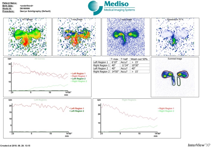
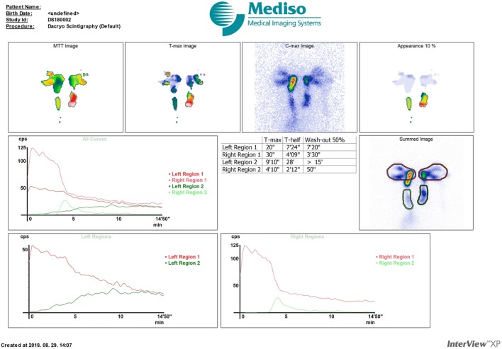
Epiphora is commonly caused by a relative or complete occlusion in the lacrimal drainage system (LDS), principally a nasolacrimal duct obstruction (NLDO). Dacryoscintigraphy (DSG), an extensively assessed imaging technique in diagnosing its abnormalities, can provide only planar images, according to which it needs to be improved. Our aim was to evaluate clinical utility of simultaneous DSG and single-photon emission computed tomography/computed tomography (SPECT/CT) combined with computed tomographic dacryocystography (CT-DCG) in the evaluation of LDS.
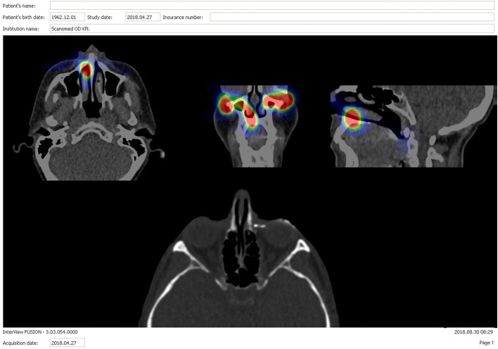
Dynamic imaging with DSG was performed, and tracer radioactivity was detected by a gamma camera. Successively, SPECT/CT images of the involved region were gained, followed by CT-DCG, during which a contrast medium was syringed into the affected LDS, and finally contrast CT scans were obtained again from the same region.
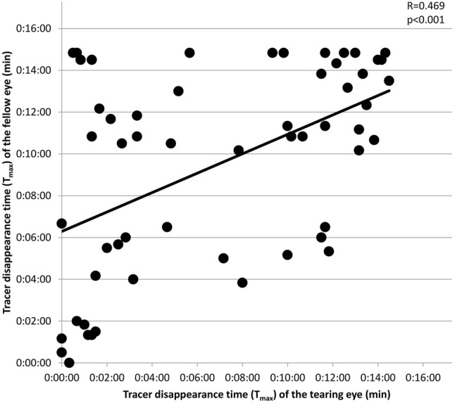
Fifty-seven patients, mean age 54.25 (± 18.26) years all with unilateral NLDO and 32 control subjects, all with patent LDS, mean age 49.88 (± 18.61) years were evaluated in the study. Delayed outflow of tearing eyes was exposed to DSG compared to the fellow and control eyes. The highest value for sensitivity was observed for SPECT/CT, followed by CT-DCG and DSG techniques, while combining DSG with SPECT/CT, DSG with CT-DCG, and SPECT/CT with CT-DCG, the sensitivity increased to 96.49%, 92.98%, and 94.73%, respectively.
Although DSG is a sensitive nuclear medicine method, it only provides useful clinical data when simultaneously supplemented with SPECT/CT and CT-DCG trials as they jointly can offer valuable information about the localization of an abnormality and verify stenosis or obstruction.
溢泪通常是由泪液引流系统(LDS)的相对或完全阻塞引起的,主要是鼻泪管阻塞(NLDO)。泪液闪烁显像(DSG)是一种广泛评估的诊断其异常的成像技术,但只能提供平面图像,因此需要改进。我们的目的是评估同时进行 DSG 和单光子发射计算机断层扫描/计算机断层扫描(SPECT/CT)结合计算机断层泪囊造影(CT-DCG)在 LDS 评估中的临床应用。
进行 DSG 动态成像,通过伽马相机检测示踪剂放射性。随后,获得受累区域的 SPECT/CT 图像,然后进行 CT-DCG,在此过程中向受累 LDS 中注入造影剂,最后再次从同一区域获得对比 CT 扫描。
研究中评估了 57 例单侧 NLDO 患者,平均年龄 54.25(±18.26)岁,以及 32 例对照受试者,所有受试者 LDS 均通畅,平均年龄 49.88(±18.61)岁。与对侧眼和对照眼相比,DSG 显示流泪眼的流出延迟。SPECT/CT 的敏感性最高,其次是 CT-DCG 和 DSG 技术,而将 DSG 与 SPECT/CT、DSG 与 CT-DCG 以及 SPECT/CT 与 CT-DCG 结合时,敏感性分别提高到 96.49%、92.98%和 94.73%。
尽管 DSG 是一种敏感的核医学方法,但只有在同时补充 SPECT/CT 和 CT-DCG 试验时,才能提供有用的临床数据,因为它们可以共同提供关于异常部位的有价值信息,并验证狭窄或阻塞。