Departments of Orthopaedics (K.W., T.G., H.W., R.M.C., and B.C.H.v.d.W.) and Anatomy (R.L.A.W.B.), University Medical Centre Utrecht, Utrecht, the Netherlands.
Department of Orthopaedics, Meander Medical Centre, Amersfoort, the Netherlands.
J Bone Joint Surg Am. 2019 Jul 17;101(14):e68. doi: 10.2106/JBJS.18.00892.
Anterior glenohumeral instability with >20% glenoid bone loss is a disorder that can be treated with the Latarjet stabilizing procedure; however, complications are common. The purposes of this study were to (1) evaluate the effect of an anatomic-specific titanium implant produced by 3-dimensional (3D) printing as a treatment option for recurrent shoulder instability with substantial glenoid bone loss and (2) compare the use of that implant with the Latarjet procedure.
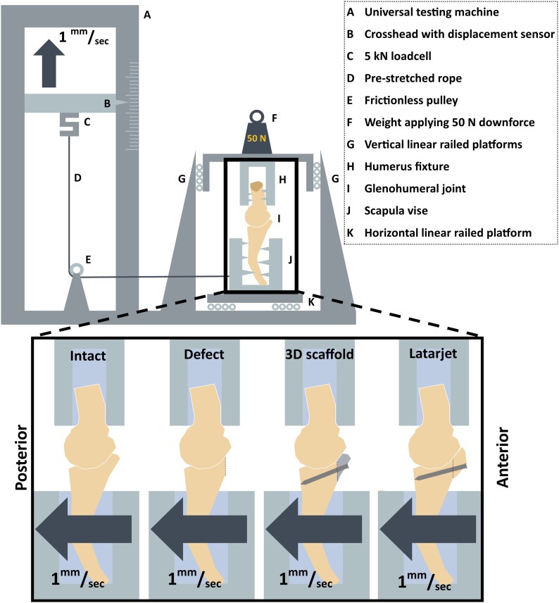
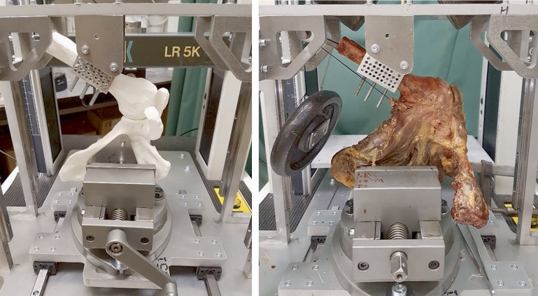
Ten fresh-frozen cadaveric shoulders (mean age at the time of death, 78 years) were tested in a biomechanical setup with the humerus in 30° of abduction and in neutral rotation. The shoulders were tested under 5 different conditions: (1) normal situation, (2) creation of an anterior glenoid defect, (3) implantation of an anatomic-specific titanium implant produced by 3D printing, and the Latarjet procedure (4) with and (5) without 10 N of load attached to the conjoined tendon. In each condition, the humerus was translated 10 mm anteriorly relative to the glenoid, and the maximum peak translational force that was necessary for this translation was measured.
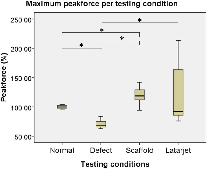
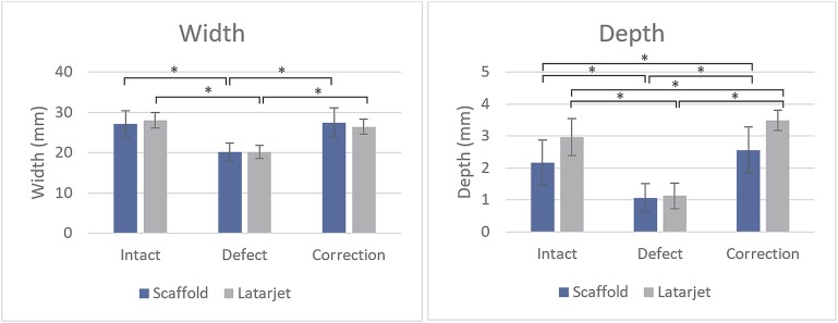
After creation of the glenoid defect, the mean translational peak force decreased by 30% ± 6% compared with that for the normal shoulder. After restoration of the original glenoid anatomy, the translational force needed to dislocate the humeral head from the glenoid significantly increased compared with that in the defect condition-to 119% ± 16% of normal (p < 0.01) with the 3D-printed anatomic-specific implant and to 121% ± 48% of normal (p < 0.01) following the Latarjet procedure. No significant differences in mean translational force were found between the anatomic-specific implant and the Latarjet procedure (p = 0.72).
The mean translational peak force needed to dislocate the humerus 10 mm anteriorly on the glenoid was higher after glenoid restoration with the 3D-printed anatomic-specific implant compared with when the glenoid had a 20% surface defect but also compared with when the glenoid was intact. No differences in mean translational peak force were found between the 3D-printed anatomic-specific glenoid implant and the Latarjet procedure, although there was less variability in the 3D-implant condition.
Novel 3D-printing technology could provide a reliable patient-specific alternative to solve problems related to traditional treatment methods for shoulder instability.
伴有>20%肩盂骨缺损的前方盂肱关节不稳定是一种可以通过 Latarjet 稳定术治疗的疾病;然而,并发症很常见。本研究的目的是:(1)评估 3D 打印的解剖特异性钛植入物作为治疗伴有大量肩盂骨缺损的复发性肩关节不稳定的治疗选择的效果;(2)比较该植入物与 Latarjet 手术的使用。
10 个新鲜冷冻的尸体肩部(死亡时的平均年龄为 78 岁)在肱骨外展 30°和中立位旋转的生物力学设置中进行测试。肩部在 5 种不同条件下进行测试:(1)正常情况,(2)前盂唇缺损的创建,(3)植入 3D 打印的解剖特异性钛植入物,(4)和(5)在未附加 10N 负荷的情况下附加到联合肌腱上。在每种情况下,肱骨头相对于盂唇向前平移 10mm,测量使这种平移所需的最大峰值平移力。
在创建盂唇缺损后,与正常肩部相比,平均平移峰值力下降了 30%±6%。在恢复原始盂唇解剖结构后,使肱骨头从盂唇脱位所需的平移力显著增加,与缺损状态相比,增加至 119%±16%(p<0.01),与 3D 打印的解剖特异性植入物相比增加至 121%±48%(p<0.01),采用 Latarjet 手术。在解剖特异性植入物和 Latarjet 手术之间,没有发现平均平移力的显著差异(p=0.72)。
在使用 3D 打印的解剖特异性植入物恢复盂唇后,使肱骨头向前平移 10mm 在盂唇上所需的平均峰值平移力高于存在 20%表面缺损的盂唇,但也高于盂唇完整的情况。在 3D 打印的解剖特异性盂唇植入物和 Latarjet 手术之间,没有发现平均峰值平移力的差异,尽管 3D 植入物的情况变化较小。
新型 3D 打印技术可以为解决与传统肩关节不稳定治疗方法相关的问题提供可靠的个体化替代方案。