Department of Radiology, Pediatric Molecular Imaging Program, Stanford University, 725 Welch Road, Stanford, CA, 94304, USA.
Department of Diagnostic and Interventional Radiology, University Medical Center of the Johannes Gutenberg-University Mainz, Mainz, Germany.
Mol Imaging Biol. 2020 Jun;22(3):722-729. doi: 10.1007/s11307-019-01409-3.
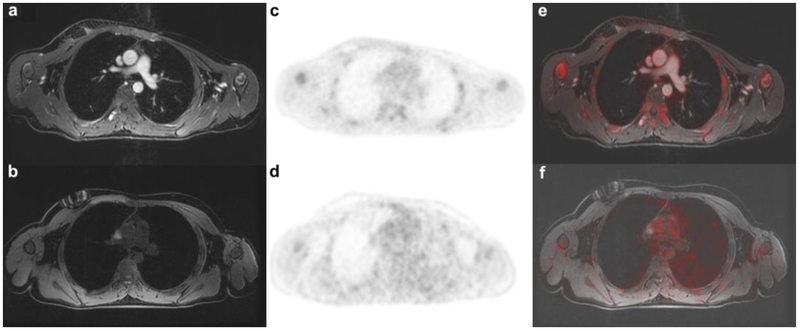
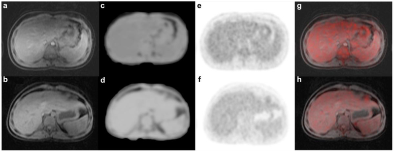
Tumor response assessments on positron emission tomography (PET)/magnetic resonance imaging (MRI) scans require correct quantification of radiotracer uptake in tumors and normal organs. Historically, MRI scans have been enhanced with gadolinium (Gd)-based contrast agents, which are now controversial due to brain deposition. Recently, ferumoxytol nanoparticles have been identified as an alternative to Gd-based contrast agents because they provide strong tissue enhancement on MR images but are not deposited in the brain. However, it is not known if the strong T1- and T2-contrast obtained with iron oxide nanoparticles such as ferumoxytol could affect MR-based attenuation correction of PET data. The purpose of our study was to investigate if ferumoxytol administration prior to a 2-deoxy-2-[F]fluoro-D-glucose [F]FDG PET/MR scan would change standardized uptake values (SUV) of normal organs.
Thirty pediatric patients (6-18 years) with malignant tumors underwent [F]FDG-PET/MR scans (dose 3 MBq/kg). Fifteen patients received an intravenous ferumoxytol injection (5 mg Fe/kg) prior to the [F]FDG-PET/MR scans (group 1). Fifteen additional age- and sex-matched patients received unenhanced [F]FDG-PET/MR scans (group 2). For attenuation correction of PET data, we used a Dixon-based gradient echo sequence (TR 4.2 ms, TE 1.1, 2.3 ms, FA 5), which accounted for soft tissue, lung, fat, and background air. We used a mixed linear effects model to compare the tissue MRI enhancement, quantified as the signal-to-noise ratio (SNR), as well as tissue radiotracer signal, quantified as SUVmean and SUVmax, between group 1 and group 2. Alpha was assumed at 0.05.
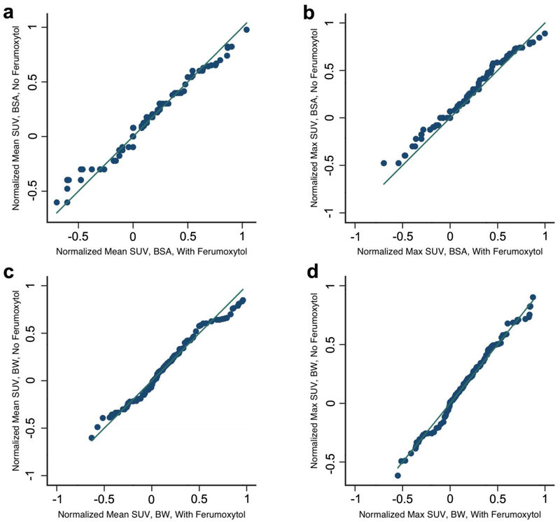
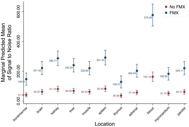
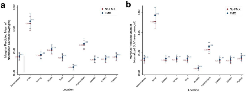
The MRI enhancement of the blood and solid extra-cerebral organs, quantified as SNR, was significantly higher on ferumoxytol-enhanced MRI scans compared to unenhanced scans (p < 0.001). However, SUVmean and SUVmax values, corrected based on the patients' body weight or body surface area, were not significantly different between the two groups (p > 0.05).
Ferumoxytol administration prior to a [F]FDG PET/MR scan did not change standardized uptake values (SUV) of solid extra-cerebral organs. This is important, because it allows injection of ferumoxytol contrast prior to a PET/MRI procedure and, thereby, significantly accelerates image acquisition times.
正电子发射断层扫描(PET)/磁共振成像(MRI)扫描上的肿瘤反应评估需要正确量化肿瘤和正常器官内的示踪剂摄取。历史上,MRI 扫描使用钆(Gd)基造影剂增强,由于脑内沉积,目前该造影剂存在争议。最近,发现氧化铁纳米颗粒(如 ferumoxytol)是 Gd 基造影剂的替代品,因为它们在 MRI 图像上提供强烈的组织增强,但不会在脑内沉积。然而,目前尚不清楚氧化铁纳米颗粒(如 ferumoxytol)获得的强 T1 和 T2 对比是否会影响基于 MRI 的 PET 数据衰减校正。我们研究的目的是调查在 2-脱氧-2-[F]氟-D-葡萄糖[F]FDG PET/MR 扫描前给予 ferumoxytol 是否会改变正常器官的标准化摄取值(SUV)。
30 名患有恶性肿瘤的儿科患者(6-18 岁)接受了[F]FDG-PET/MR 扫描(剂量 3MBq/kg)。15 名患者在[F]FDG-PET/MR 扫描前接受静脉注射 ferumoxytol(5mgFe/kg)(第 1 组)。另外 15 名年龄和性别匹配的患者接受了未经增强的[F]FDG-PET/MR 扫描(第 2 组)。为了对 PET 数据进行衰减校正,我们使用了基于 Dixon 的梯度回波序列(TR 4.2ms,TE 1.1、2.3ms,FA 5),该序列考虑了软组织、肺、脂肪和背景空气。我们使用混合线性效应模型比较了第 1 组和第 2 组之间的组织 MRI 增强,用信噪比(SNR)表示,以及组织示踪剂信号,用 SUVmean 和 SUVmax 表示。假设α值为 0.05。
与未增强的 MRI 扫描相比,血液和实体外脑器官的 MRI 增强(用 SNR 表示)在 ferumoxytol 增强 MRI 扫描中明显更高(p<0.001)。然而,两组之间基于患者体重或体表面积校正的 SUVmean 和 SUVmax 值无显著差异(p>0.05)。
在[F]FDG PET/MR 扫描前给予 ferumoxytol 不会改变实体外脑器官的标准化摄取值(SUV)。这很重要,因为它允许在 PET/MRI 程序前注射 ferumoxytol 造影剂,从而显著加快图像采集时间。