Department of Obstetrics and Gynecology, Korea University College of Medicine, Seoul, Korea.
Department of Pathology, Korea University College of Medicine, Seoul, Korea.
J Gynecol Oncol. 2019 Sep;30(5):e75. doi: 10.3802/jgo.2019.30.e75.
Human epidermal growth factor receptor-2 (HER2) and 3 (HER3) belong to the epidermal growth factor receptor (EGFR) family of transmembrane receptor tyrosine kinases. In this study, we assessed HER2/HER3 expression levels in specimens of epithelial ovarian cancer and determined their correlation with clinical features of ovarian cancer.
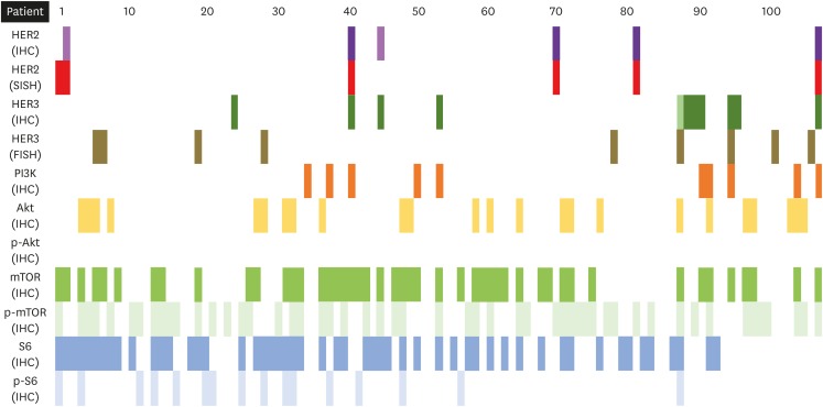
Tissue microarrays (TMAs) were prepared from paraffin blocks of 105 ovarian tumour samples. HER2, HER3, PI3K, Akt, p-Akt, mTOR, p-mTOR, S6, and p-S6 expression levels were investigated using immunohistochemistry (IHC). and amplifications were determined using in situ hybridization (ISH). The correlation between HER2/3 expression and disease outcome of the patients including surgical outcome, progression-free survival (PFS) and overall survival (OS) was analysed.
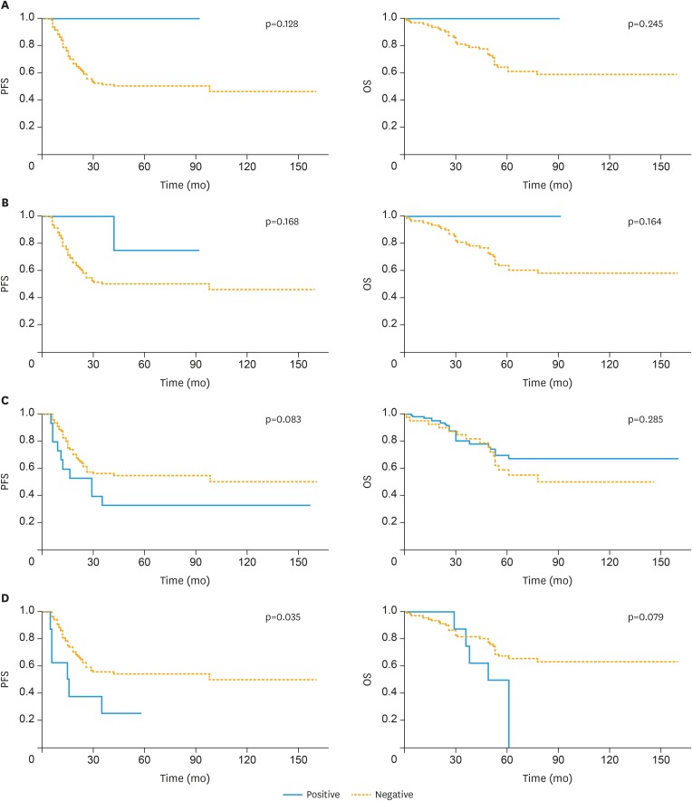
HER2 positivity was 3.8% by IHC and 5.7% by ISH, whereas that of HER3 was 12.4% and 8.6%, respectively. HER2 status by either IHC or ISH was not related to PFS (p=0.128, 0.168, respectively) and OS (p=0.245, 0.164, respectively). However, the HER3 status determined using fluorescence ISH was associated with poor PFS (p=0.035 on log rank test), which was a significant risk factor even after adjusting other possible risk factors in multivariate analysis (hazard ratio=2.377 [1.18-7.49], p=0.021). Expressions of Akt, p-mTOR, and S6 were also related with poor progression (p=0.008, 0.049, 0.014, respectively).
HER3 is possibly an independent marker for poor prognosis in individuals with ovarian cancer, as the HER3 signalling pathway is distinct from that of HER2. The possibility of targeted therapy for patients with alteration in ovarian cancer should be evaluated.
人类表皮生长因子受体 2(HER2)和 3(HER3)属于表皮生长因子受体(EGFR)家族的跨膜受体酪氨酸激酶。本研究评估了上皮性卵巢癌标本中 HER2/HER3 的表达水平,并确定了它们与卵巢癌临床特征的相关性。
从 105 例卵巢肿瘤样本的石蜡块中制备组织微阵列(TMA)。使用免疫组织化学(IHC)检测 HER2、HER3、PI3K、Akt、p-Akt、mTOR、p-mTOR、S6 和 p-S6 的表达水平。使用原位杂交(ISH)确定 HER2/3 扩增。分析 HER2/3 表达与患者疾病结局(包括手术结局、无进展生存期(PFS)和总生存期(OS))的相关性。
IHC 检测的 HER2 阳性率为 3.8%,ISH 检测的阳性率为 5.7%,而 HER3 的阳性率分别为 12.4%和 8.6%。无论是 IHC 还是 ISH 检测的 HER2 状态均与 PFS(p=0.128,0.168)和 OS(p=0.245,0.164)无关。然而,荧光 ISH 检测的 HER3 状态与不良 PFS 相关(对数秩检验,p=0.035),即使在多变量分析中调整其他可能的风险因素后,这也是一个显著的风险因素(风险比=2.377[1.18-7.49],p=0.021)。Akt、p-mTOR 和 S6 的表达也与进展不良相关(p=0.008,0.049,0.014)。
HER3 可能是卵巢癌患者不良预后的独立标志物,因为 HER3 信号通路与 HER2 不同。应该评估卵巢癌患者改变后进行靶向治疗的可能性。