• 文献检索
  • 文档翻译
  • 深度研究
  • 学术资讯
  • Suppr Zotero 插件Zotero 插件
  • 邀请有礼
  • 套餐&价格
  • 历史记录
应用&插件
Suppr Zotero 插件Zotero 插件浏览器插件Mac 客户端Windows 客户端微信小程序
定价
高级版会员购买积分包购买API积分包
服务
文献检索文档翻译深度研究API 文档MCP 服务
关于我们
关于 Suppr公司介绍联系我们用户协议隐私条款
关注我们

Suppr 超能文献

核心技术专利:CN118964589B侵权必究
粤ICP备2023148730 号-1Suppr @ 2026

文献检索

告别复杂PubMed语法,用中文像聊天一样搜索,搜遍4000万医学文献。AI智能推荐,让科研检索更轻松。

立即免费搜索

文件翻译

保留排版,准确专业,支持PDF/Word/PPT等文件格式,支持 12+语言互译。

免费翻译文档

深度研究

AI帮你快速写综述,25分钟生成高质量综述,智能提取关键信息,辅助科研写作。

立即免费体验

在中国,光学相干断层扫描(OCT)图像中的高反射灶作为糖尿病性黄斑水肿患者接受康柏西普治疗预后不良的生物标志物。

Hyperreflective foci in OCT image as a biomarker of poor prognosis in diabetic macular edema patients treating with Conbercept in China.

作者信息

Liu Shulin, Wang Desai, Chen Fei, Zhang Xuedong

机构信息

The First Affiliated Hospital of Chongqing Medical University, Chongqing Key Laboratory of Ophthalmology and Chongqing Eye Institute, 1 You Yi Road, Yu Zhong District, Chongqing, 400016, People's Republic of China.

Ophthalmology Department, The people's Hospital of BiShan District of Chongqing City, Chongqing, People's Republic of China.

出版信息

BMC Ophthalmol. 2019 Jul 23;19(1):157. doi: 10.1186/s12886-019-1168-0.

DOI:10.1186/s12886-019-1168-0
PMID:31337360
原文链接:https://pmc.ncbi.nlm.nih.gov/articles/PMC6651859/
Abstract

PURPOSE

To investigate the dynamic changes of hyperreflective foci (HF) in diabetic macular edema (DME) patients during the intravitreal Conbercept treatment in China.

METHODS

DME Patients receiving intravitreal Conbercept (IVC) injections during the year 2016-2017 were retrospectively investigated. Thirteen patients (26 eyes) were recruited in this study. They received IVC once a month for 3 consecutive months. The number and location of HFs, the best-corrected visual acuity (BCVA) and central macular thickness (CMT) at each visit were analyzed and compared.

RESULTS

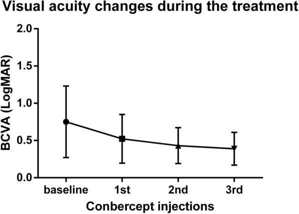
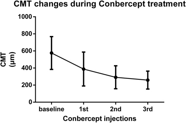
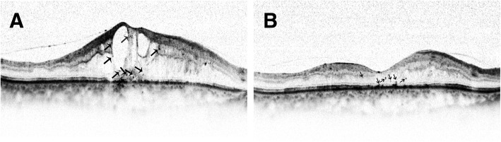
After the first injection, BCVA (LogMAR) was increased from 0.75 ± 0.48 to 0.43 ± 0.24 (p < 0.05), CMT improved from 575.9 ± 191.9 to 388.2 ± 198.5 μm (p = 0.014). However, the BCVA and CMT had no statistical difference after the second and third injection as compared with those after the first injection respectively. The baseline number of HFs was 5.39 ± 4.24, 5.15 ± 5.17 and 0.88 ± 1.90 in the inner retinal, outer retinal and subretinal layer respectively. The number of HFs in these three retinal layers decreased significantly after the first injection (p = 0.0045, p < 0.0001 and p = 0.0045, respectively). However, after the second injection, only the number of HFs in the inner retinal layer experienced a further decrease. After the third injection, no statistically significant HFs changes was observed in each retinal layers. Correlation analysis showed that there was a positive significant correlation between the baseline number of HFs in the inner retina, outer retina, subretina and final BCVA (r = 0.571, p = 0.002; r = 0.464, p = 0.017; r = 0.405, p = 0.04 respectively). There was also a significant positive correlation between outer retinal HFs reduction, total retinal HFs reduction and increase of BCVA (r = 0.40, p = 0.043 and r = 0.393, p = 0.04 respectively). There were no severe ocular adverse reactions or systemic adverse events.

CONCLUSIONS

Conbercept is effective and safe in the treatment of DME. HFs can act as a biomarker of poor final visual outcome.

摘要

目的

研究中国糖尿病性黄斑水肿(DME)患者玻璃体内注射康柏西普治疗期间高反射灶(HF)的动态变化。

方法

回顾性研究2016年至2017年期间接受玻璃体内注射康柏西普(IVC)的DME患者。本研究招募了13例患者(26只眼)。他们连续3个月每月接受一次IVC注射。分析并比较每次就诊时HF的数量和位置、最佳矫正视力(BCVA)和中心黄斑厚度(CMT)。

结果

首次注射后,BCVA(LogMAR)从0.75±0.48提高到0.43±0.24(p<0.05),CMT从575.9±191.9改善至388.2±198.5μm(p = 0.014)。然而,第二次和第三次注射后的BCVA和CMT与首次注射后相比分别无统计学差异。HF的基线数量在内视网膜、外视网膜和视网膜下分别为5.39±4.24、5.15±5.17和0.88±1.90。首次注射后这三个视网膜层的HF数量均显著减少(分别为p = 0.0045、p<0.0001和p = 0.0045)。然而,第二次注射后,仅内视网膜层的HF数量进一步减少。第三次注射后,各视网膜层未观察到HF有统计学意义的变化。相关性分析显示,内视网膜、外视网膜、视网膜下HF的基线数量与最终BCVA之间存在显著正相关(r = 0.571,p = 0.002;r = 0.464,p = 0.017;r = 0.405,p = 0.04)。外视网膜HF减少、总视网膜HF减少与BCVA增加之间也存在显著正相关(r = 0.40,p = 0.043和r = 0.393,p = 0.04)。未出现严重眼部不良反应或全身不良事件。

结论

康柏西普治疗DME有效且安全。HF可作为最终视力不佳的生物标志物。

https://cdn.ncbi.nlm.nih.gov/pmc/blobs/7cef/6651859/1a2980ed71cb/12886_2019_1168_Fig4_HTML.jpg
https://cdn.ncbi.nlm.nih.gov/pmc/blobs/7cef/6651859/d4ca57c2ba05/12886_2019_1168_Fig1_HTML.jpg
https://cdn.ncbi.nlm.nih.gov/pmc/blobs/7cef/6651859/58cc202e294e/12886_2019_1168_Fig2_HTML.jpg
https://cdn.ncbi.nlm.nih.gov/pmc/blobs/7cef/6651859/e78e6a6fc770/12886_2019_1168_Fig3_HTML.jpg
https://cdn.ncbi.nlm.nih.gov/pmc/blobs/7cef/6651859/1a2980ed71cb/12886_2019_1168_Fig4_HTML.jpg

相似文献

1
Hyperreflective foci in OCT image as a biomarker of poor prognosis in diabetic macular edema patients treating with Conbercept in China.在中国,光学相干断层扫描(OCT)图像中的高反射灶作为糖尿病性黄斑水肿患者接受康柏西普治疗预后不良的生物标志物。
BMC Ophthalmol. 2019 Jul 23;19(1):157. doi: 10.1186/s12886-019-1168-0.
2
[Treatment response to Conbercept of different types of diabetic macular edema classified based on optical coherence tomography].[基于光学相干断层扫描分类的不同类型糖尿病性黄斑水肿对康柏西普的治疗反应]
Nan Fang Yi Ke Da Xue Xue Bao. 2021 Oct 20;41(10):1501-1508. doi: 10.12122/j.issn.1673-4254.2021.10.08.
3
Correlation of retinal layer changes with vision gain in diabetic macular edema during conbercept treatment.康柏西普治疗糖尿病性黄斑水肿期间视网膜层变化与视力改善的相关性
BMC Ophthalmol. 2019 May 31;19(1):123. doi: 10.1186/s12886-019-1131-0.
4
Comparison of 12-month therapeutic effect of conbercept and ranibizumab for diabetic macular edema: a real-life clinical practice study.康柏西普与雷珠单抗治疗糖尿病性黄斑水肿12个月的疗效比较:一项真实世界临床实践研究
BMC Ophthalmol. 2017 Aug 25;17(1):158. doi: 10.1186/s12886-017-0554-8.
5
CORRELATION OF OPTICAL COHERENCE TOMOGRAPHIC HYPERREFLECTIVE FOCI WITH VISUAL OUTCOMES IN DIFFERENT PATTERNS OF DIABETIC MACULAR EDEMA.不同模式糖尿病黄斑水肿中光学相干断层扫描高反射灶与视力预后的相关性
Retina. 2016 Sep;36(9):1630-9. doi: 10.1097/IAE.0000000000000995.
6
One-Year Outcome of Conbercept Therapy for Diabetic Macular Edema.康柏西普治疗糖尿病性黄斑水肿的一年疗效
Curr Eye Res. 2018 Feb;43(2):218-223. doi: 10.1080/02713683.2017.1379542. Epub 2017 Dec 21.
7
OPTICAL COHERENCE TOMOGRAPHY PREDICTORS OF SHORT-TERM VISUAL ACUITY IN EYES WITH MACULAR EDEMA SECONDARY TO RETINAL VEIN OCCLUSION TREATED WITH INTRAVITREAL CONBERCEPT.玻璃体内注射康柏西普治疗视网膜静脉阻塞继发黄斑水肿患者短期视力的光学相干断层扫描预测指标
Retina. 2020 Apr;40(4):773-785. doi: 10.1097/IAE.0000000000002444.
8
One-year outcomes of novel VEGF decoy receptor therapy with intravitreal conbercept in diabetic retinopathy-induced macular edema.玻璃体内注射康柏西普新型VEGF诱饵受体疗法治疗糖尿病性视网膜病变引起的黄斑水肿的一年疗效
Mol Vis. 2019 Oct 21;25:636-644. eCollection 2019.
9
MEASUREMENTS OF RETINAL FLUID BY OPTICAL COHERENCE TOMOGRAPHY LEAKAGE IN DIABETIC MACULAR EDEMA: A Biomarker of Visual Acuity Response to Treatment.光学相干断层扫描渗漏测量在糖尿病性黄斑水肿中的应用:视力反应治疗的生物标志物。
Retina. 2019 Jan;39(1):52-60. doi: 10.1097/IAE.0000000000001905.
10
Evaluation of inner and outer retinal thickness in patients receiving intravitreal ranibizumab injections for diabetic macular edema.接受玻璃体内注射雷珠单抗治疗糖尿病性黄斑水肿患者的视网膜内层和外层厚度评估。
Eur J Ophthalmol. 2016 Jan-Feb;26(1):48-53. doi: 10.5301/ejo.5000655. Epub 2015 Jul 15.

引用本文的文献

1
Effect of Faricimab versus Aflibercept on Hyperreflective Foci in Patients with Diabetic Macular Edema from the YOSEMITE/RHINE Trials.在YOSEMITE/RHINE试验中,法西单抗与阿柏西普对糖尿病性黄斑水肿患者高反射灶的影响。
Ophthalmol Sci. 2025 Apr 19;5(5):100798. doi: 10.1016/j.xops.2025.100798. eCollection 2025 Sep-Oct.
2
Hyperreflective Retinal Foci (HRF): Definition and Role of an Invaluable OCT Sign.高反射性视网膜病灶(HRF):一种重要的光学相干断层扫描(OCT)征象的定义及作用
J Clin Med. 2025 Apr 27;14(9):3021. doi: 10.3390/jcm14093021.
3
[MiHUBx: a digital progress hub for the use of intersectoral clinical data sets using the example of diabetic macular edema].

本文引用的文献

1
Correlation of retinal layer changes with vision gain in diabetic macular edema during conbercept treatment.康柏西普治疗糖尿病性黄斑水肿期间视网膜层变化与视力改善的相关性
BMC Ophthalmol. 2019 May 31;19(1):123. doi: 10.1186/s12886-019-1131-0.
2
Number of Hyperreflective Foci in the Outer Retina Correlates with Inflammation and Photoreceptor Degeneration in Retinitis Pigmentosa.视网膜色素变性中外层视网膜高反射灶的数量与炎症和光感受器变性相关。
Ophthalmol Retina. 2018 Jul;2(7):726-734. doi: 10.1016/j.oret.2017.07.020. Epub 2017 Oct 10.
3
SHORT-TERM EFFICACY OF CONBERCEPT AND RANIBIZUMAB FOR POLYPOIDAL CHOROIDAL VASCULOPATHY.
[MiHUBx:以糖尿病性黄斑水肿为例的跨部门临床数据集使用数字进展中心]
Ophthalmologie. 2025 Apr;122(4):262-269. doi: 10.1007/s00347-024-02146-x. Epub 2024 Dec 18.
4
A spectral domain optical coherence tomography (SD-OCT) study of intraretinal hyperreflective foci and its relationship with visual acuity and grades of retinopathy among patients of diabetes mellitus in Indian population.印度人群糖尿病患者视网膜内高反射灶的光谱域光学相干断层扫描(SD-OCT)研究及其与视力和视网膜病变分级的关系。
Med J Armed Forces India. 2024 Sep-Oct;80(5):572-577. doi: 10.1016/j.mjafi.2024.04.019. Epub 2024 Jul 3.
5
Sex Differences in Inflammation-Related Biomarkers Detected with OCT in Patients with Diabetic Macular Edema.糖尿病性黄斑水肿患者中通过光学相干断层扫描检测到的炎症相关生物标志物的性别差异。
Ophthalmol Sci. 2024 Jul 18;4(6):100580. doi: 10.1016/j.xops.2024.100580. eCollection 2024 Nov-Dec.
6
Intraretinal Hyper-Reflective Foci Are Almost Universally Present and Co-Localize With Intraretinal Fluid in Diabetic Macular Edema.视网膜内高反射焦点几乎普遍存在,并与糖尿病性黄斑水肿的视网膜内液共存。
Invest Ophthalmol Vis Sci. 2024 May 1;65(5):26. doi: 10.1167/iovs.65.5.26.
7
An Optical Coherence Tomography-Based Measure as an Independent Estimate of Retinal Function in Retinitis Pigmentosa.一种基于光学相干断层扫描的测量方法作为视网膜色素变性视网膜功能的独立评估指标。
Diagnostics (Basel). 2023 Nov 24;13(23):3521. doi: 10.3390/diagnostics13233521.
8
Automated evaluation of retinal hyperreflective foci changes in diabetic macular edema patients before and after intravitreal injection.糖尿病性黄斑水肿患者玻璃体内注射前后视网膜高反射灶变化的自动评估
Front Med (Lausanne). 2023 Oct 6;10:1280714. doi: 10.3389/fmed.2023.1280714. eCollection 2023.
9
Evaluation of the efficacy of anti-vascular endothelial growth factors in diabetic macular edema with retinal inner and outer layers disorganization.评估抗血管内皮生长因子治疗视网膜内、外层组织紊乱性糖尿病黄斑水肿的疗效。
Acta Diabetol. 2023 Oct;60(10):1391-1398. doi: 10.1007/s00592-023-02121-z. Epub 2023 Jun 28.
10
Automated Region of Interest Selection Improves Deep Learning-Based Segmentation of Hyper-Reflective Foci in Optical Coherence Tomography Images.自动感兴趣区域选择可改善光学相干断层扫描图像中基于深度学习的高反射灶分割。
J Clin Med. 2022 Dec 14;11(24):7404. doi: 10.3390/jcm11247404.
康柏西普和雷珠单抗治疗息肉状脉络膜血管病变的短期疗效。
Retina. 2019 May;39(5):889-895. doi: 10.1097/IAE.0000000000002035.
4
One-Year Outcome of Conbercept Therapy for Diabetic Macular Edema.康柏西普治疗糖尿病性黄斑水肿的一年疗效
Curr Eye Res. 2018 Feb;43(2):218-223. doi: 10.1080/02713683.2017.1379542. Epub 2017 Dec 21.
5
Comparison of 12-month therapeutic effect of conbercept and ranibizumab for diabetic macular edema: a real-life clinical practice study.康柏西普与雷珠单抗治疗糖尿病性黄斑水肿12个月的疗效比较:一项真实世界临床实践研究
BMC Ophthalmol. 2017 Aug 25;17(1):158. doi: 10.1186/s12886-017-0554-8.
6
SHORT-TERM OUTCOMES AFTER INTRAVITREAL INJECTIONS OF CONBERCEPT VERSUS RANIBIZUMAB FOR THE TREATMENT OF RETINOPATHY OF PREMATURITY.康柏西普玻璃体腔内注射与雷珠单抗治疗早产儿视网膜病变的短期疗效比较。
Retina. 2018 Aug;38(8):1595-1604. doi: 10.1097/IAE.0000000000001763.
7
Efficacy and safety of conbercept as a primary treatment for choroidal neovascularization secondary to punctate inner choroidopathy.康柏西普作为点状内层脉络膜病变继发脉络膜新生血管主要治疗方法的疗效和安全性。
BMC Ophthalmol. 2017 Jun 12;17(1):87. doi: 10.1186/s12886-017-0481-8.
8
Evaluation of hyperreflective foci as a prognostic factor of visual outcome in retinal vein occlusion.评估高反射灶作为视网膜静脉阻塞视力预后的一个因素。
Int J Ophthalmol. 2017 Apr 18;10(4):605-612. doi: 10.18240/ijo.2017.04.17. eCollection 2017.
9
Comparison of Conbercept with Ranibizumab for the Treatment of Macular Edema Secondary to Branch Retinal Vein Occlusion.康柏西普与雷珠单抗治疗视网膜分支静脉阻塞继发黄斑水肿的比较
Curr Eye Res. 2017 Aug;42(8):1174-1178. doi: 10.1080/02713683.2017.1285943. Epub 2017 Apr 25.
10
HYPERREFLECTIVE FOCI AS AN INDEPENDENT VISUAL OUTCOME PREDICTOR IN MACULAR EDEMA DUE TO RETINAL VASCULAR DISEASES TREATED WITH INTRAVITREAL DEXAMETHASONE OR RANIBIZUMAB.高反射灶作为玻璃体内注射地塞米松或雷珠单抗治疗的视网膜血管疾病所致黄斑水肿的独立视觉预后预测指标
Retina. 2016 Dec;36(12):2319-2328. doi: 10.1097/IAE.0000000000001070.