Sorby-Adams Annabel J, Leonard Anna V, Elms Levi E, Marian Oana C, Hoving Jan W, Yassi Nawaf, Vink Robert, Thornton Emma, Turner Renée J
Adelaide Medical School, Adelaide Centre for Neuroscience Research, The University of Adelaide, Adelaide, SA, Australia.
Department of Medicine and Neurology, Melbourne Brain Centre at the Royal Melbourne Hospital, University of Melbourne, Melbourne, VIC, Australia.
Front Neurosci. 2019 Jul 9;13:587. doi: 10.3389/fnins.2019.00587. eCollection 2019.
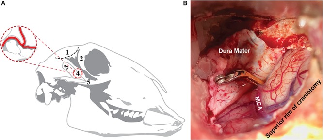
Cerebral edema and elevated intracranial pressure (ICP) are the leading cause of death in the first week following stroke. Despite this, current treatments are limited and fail to address the underlying mechanisms of swelling, highlighting the need for targeted treatments. When screening promising novel agents, it is essential to use clinically relevant large animal models to increase the likelihood of successful clinical translation. As such, we sought to develop a survival model of transient middle cerebral artery occlusion (tMCAO) in the sheep and subsequently characterize the temporal profile of cerebral edema and elevated ICP following stroke in this novel, clinically relevant model.
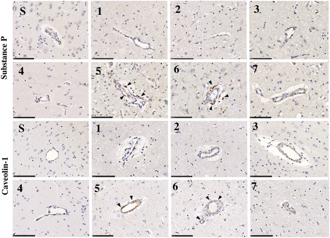
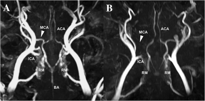
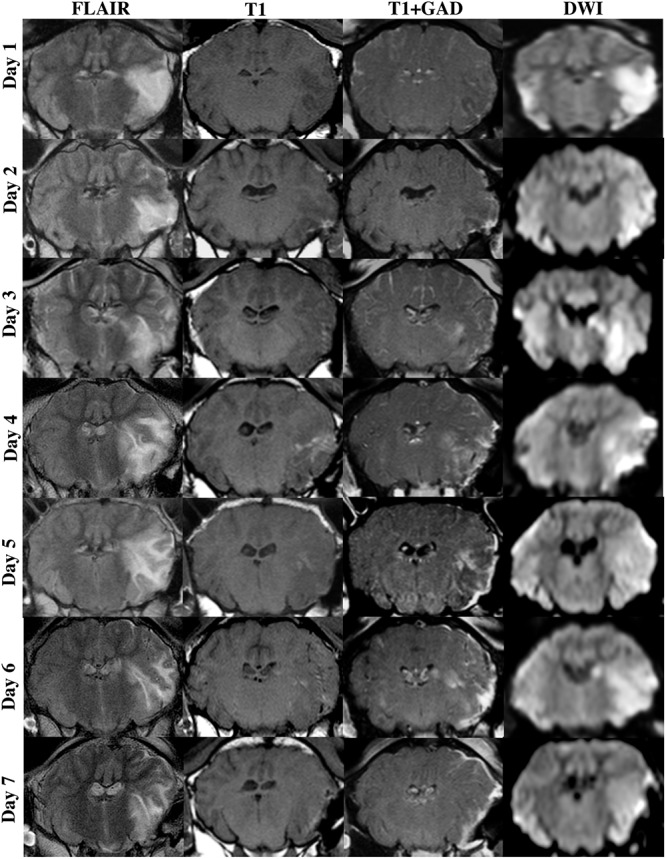
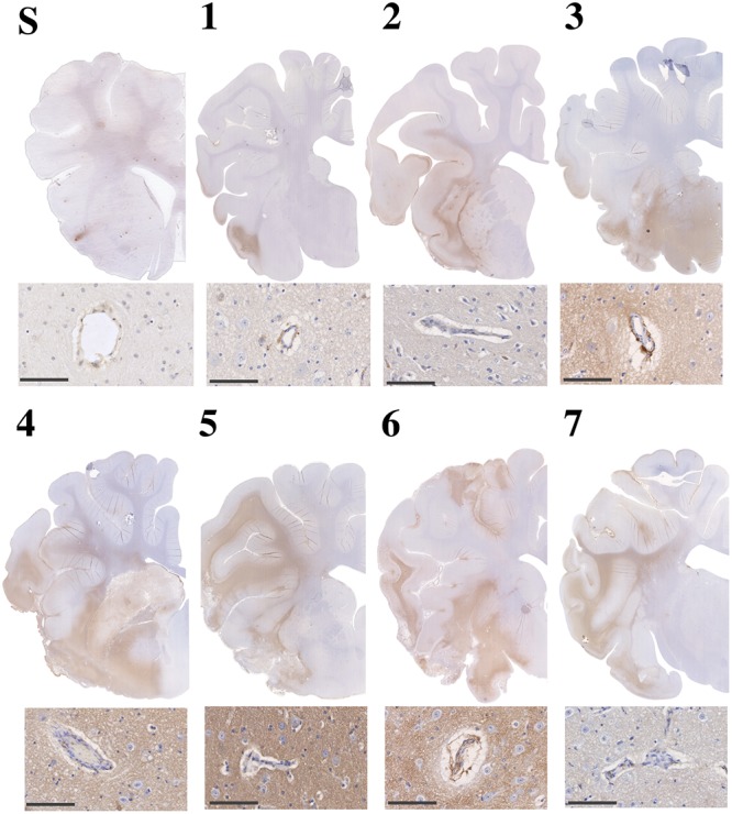
Merino-sheep (27M;31F) were anesthetized and subject to 2 h tMCAO with reperfusion or sham surgery. Following surgery, animals were allowed to recover and returned to their home pens. At preselected times points ranging from 1 to 7 days post-stroke, animals were re-anesthetized, ICP measured for 4 h, followed by imaging with MRI to determine cerebral edema, midline shift and infarct volume (FLAIR, T2 and DWI). Animals were subsequently euthanized and their brain removed for immunohistochemical analysis. Serum and cerebrospinal fluid samples were also collected and analyzed for substance P (SP) using ELISA.
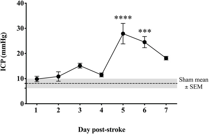
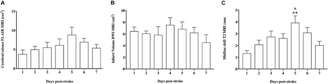
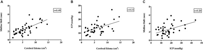
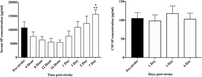
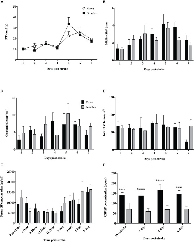
Intracranial pressure and MRI scans were normal in sham animals. Following stroke, ICP rose gradually over time and by 5 days was significantly ( < 0.0001) elevated above sham levels. Profound cerebral edema was observed as early as 2 days post-stroke and continued to evolve out to 6 days, resulting in significant midline shift which was most prominent at 5 days post-stroke ( < 0.01), in keeping with increasing ICP. Serum SP levels were significantly elevated ( < 0.01) by 7 days post-tMCAO.
We have successfully developed a survival model of ovine tMCAO and characterized the temporal profile of ICP. Peak ICP elevation, cerebral edema and midline shift occurred at days 5-6 following stroke, accompanied by an elevation in serum SP. Our findings suggest that novel therapeutic agents screened in this model targeting cerebral edema and elevated ICP would most likely be effective when administered prior to 5 days, or as early as possible following stroke onset.
脑水肿和颅内压(ICP)升高是卒中后第一周内的主要死亡原因。尽管如此,目前的治疗方法有限,未能解决肿胀的潜在机制,这凸显了靶向治疗的必要性。在筛选有前景的新型药物时,使用临床相关的大型动物模型以增加成功临床转化的可能性至关重要。因此,我们试图建立绵羊短暂性大脑中动脉闭塞(tMCAO)的存活模型,并随后在这个新型的、临床相关的模型中描述卒中后脑水肿和ICP升高的时间进程。
对美利奴绵羊(27只雄性;31只雌性)进行麻醉,实施2小时的tMCAO并进行再灌注或假手术。手术后,让动物恢复并放回它们的畜栏。在卒中后1至7天的预选时间点,再次对动物进行麻醉,测量ICP 4小时,随后进行MRI成像以确定脑水肿、中线移位和梗死体积(液体衰减反转恢复序列、T2加权像和弥散加权成像)。随后对动物实施安乐死并取出其大脑进行免疫组织化学分析。还收集血清和脑脊液样本,并用酶联免疫吸附测定法(ELISA)分析P物质(SP)。
假手术动物的颅内压和MRI扫描结果正常。卒中后,ICP随时间逐渐升高,到第5天时显著(<0.0001)高于假手术水平。早在卒中后2天就观察到严重的脑水肿,并持续发展至6天,导致明显的中线移位,在卒中后第5天最为明显(<0.01),这与ICP升高一致。tMCAO后7天血清SP水平显著升高(<0.01)。
我们成功建立了绵羊tMCAO的存活模型,并描述了ICP的时间进程。卒中后第5至6天出现ICP升高峰值、脑水肿和中线移位,并伴有血清SP升高。我们的研究结果表明,在该模型中筛选的针对脑水肿和ICP升高的新型治疗药物,在卒中后5天之前或尽可能早地给药时最有可能有效。