Chen Gang, Luo Xiaohe, Wang Wenjin, Wang Yimei, Zhu Fei, Wang Wei
Department of Plastic and Reconstructive Surgery, Shanghai Ninth People's Hospital, Shanghai Jiao Tong University School of Medicine, Shanghai, China.
Department of Plastic Surgery, First Affiliated Hospital of Anhui Medical University, Hefei, China.
Front Cell Neurosci. 2019 Jul 9;13:304. doi: 10.3389/fncel.2019.00304. eCollection 2019.
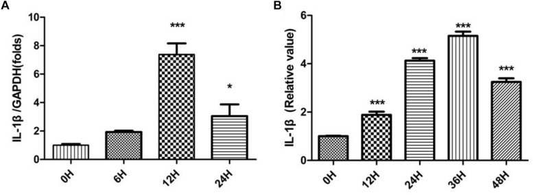
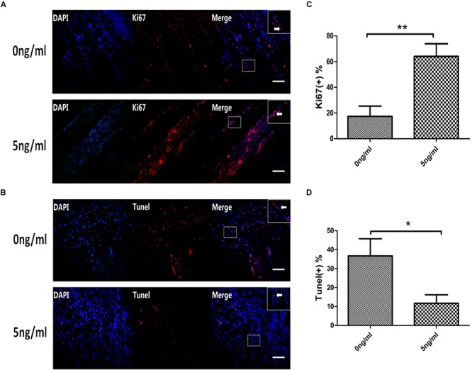
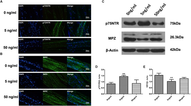
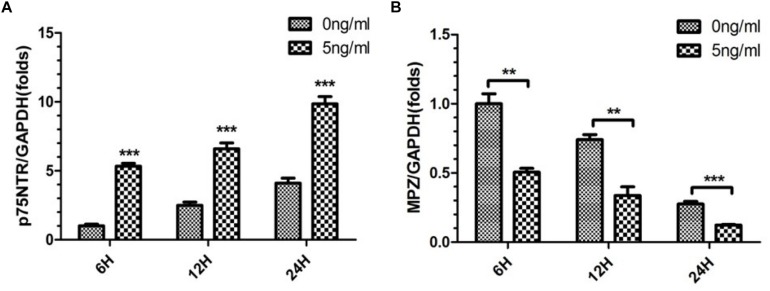
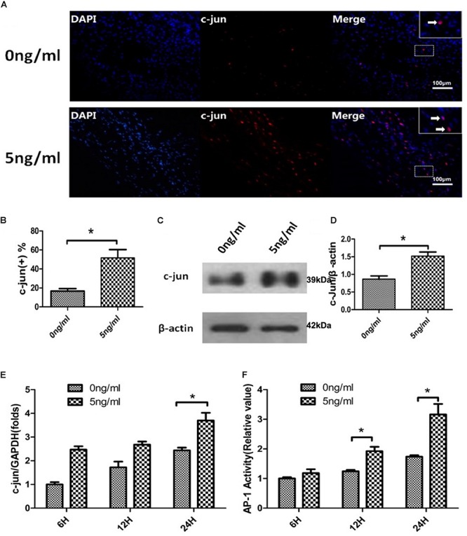
Schwann cells (SCs) de-differentiate in Wallerian degeneration (WD) following nerve injury and, by doing so, can actively promote nerve repair and functional recovery. An innate-immune response is an important component of the complex of events referred to as WD. Damaged peripheral nervous system SCs produce IL-1β and other inflammatory cytokines. We hypothesized that, in addition to a role in immune responses, IL-1β participates in de-differentiation and proliferation of SCs. qPCR and ELISA demonstrated that expression of IL-1β mRNAs and protein increased after nerve injury. Immunofluorescent staining and western blotting demonstrated that expression of the p75 neurotrophin receptor (p75NTR) was significantly increased and levels of myelin protein zero (MPZ) were significantly decreased after IL-1β exposure compared with control groups WD. Additionally, qPCR demonstrated that IL-1β elevated expression of the de-differentiation gene p75NTR and decreased expression of myelination locus MPZ and promoted SCs de-differentiation. Furthermore, immunofluorescent staining, western blotting, qPCR and ELISA revealed that IL-1β promoted c-JUN expression and activation of AP-1 activity of SCs in an WD model. Finally, Immunofluorescent staining illustrated that IL-1β elevated expression of Ki67 in SCs nuclei, the apoptosis of SCs were detected by TUNEL. SCs of WD produce IL-1β which promotes SCs de-differentiation and proliferation.
在神经损伤后的沃勒变性(WD)过程中,施万细胞(SCs)会去分化,并且通过这种方式能够积极促进神经修复和功能恢复。先天免疫反应是被称为WD的一系列复杂事件的重要组成部分。受损的外周神经系统SCs会产生白细胞介素-1β(IL-1β)和其他炎性细胞因子。我们推测,IL-1β除了在免疫反应中发挥作用外,还参与SCs的去分化和增殖。定量聚合酶链反应(qPCR)和酶联免疫吸附测定(ELISA)表明,神经损伤后IL-1β mRNA和蛋白的表达增加。免疫荧光染色和蛋白质印迹法表明,与对照组WD相比,IL-1β暴露后p75神经营养因子受体(p75NTR)的表达显著增加,髓磷脂蛋白零(MPZ)的水平显著降低。此外,qPCR表明IL-1β提高了去分化基因p75NTR的表达,降低了髓鞘形成位点MPZ的表达,并促进SCs去分化。此外,免疫荧光染色、蛋白质印迹法、qPCR和ELISA显示,在WD模型中,IL-1β促进SCs中c-JUN的表达和AP-1活性的激活。最后,免疫荧光染色表明IL-1β提高了SCs细胞核中Ki67的表达,通过末端脱氧核苷酸转移酶介导的缺口末端标记法(TUNEL)检测SCs的凋亡。WD的SCs产生IL-1β,其促进SCs去分化和增殖。