Berg Arthur, Voigt Brett, Kaul Sanjeev
Department of Trauma and Surgical Critical Care, Hackensack Meridian Health Center, Hackensack, NJ, USA.
Department of Surgery, Hackensack Meridian Health Center, Hackensack, NJ, USA.
Trauma Case Rep. 2019 Jun 27;22:100193. doi: 10.1016/j.tcr.2019.100193. eCollection 2019 Aug.
Historically, in the pediatric population, there is a highly selective approach for repeat imaging given the risk of radiation and costs. In the lieu of this, frequent neurological checks and even ICP monitoring has been used as an adjunct, although not always successful. We present a case of a pediatric patient with a late evolving epidural hematoma in the setting of a depressed skull fracture, and present an argument for serial CT imaging in a select patient population similar to his.
Discuss the unique presentation, diagnosis, and management of an expanding epidural hematoma in a pediatric patient with a depressed skull fracture and the need for aggressive repeat imaging in this setting.
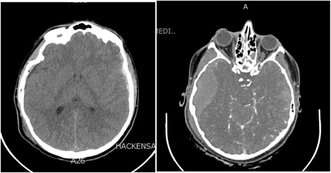
Patient is a 15-year-old boy who presented to our trauma bay after being the victim of a hit and run while skateboarding. His injuries included a depressed comminuted skull fracture and bilateral SDH. Additionally, a stat CT angiogram was obtained due to a basilar skull fracture. A rapidly evolving EDH with impending herniation was found, which was nearly fatal and was not present on the initial CT scan. He required emergent evacuation with a hemi-craniectomy where he was found to have a laceration of his dural vessels as well as his middle meningeal artery. Post operatively he did well and regained full neurologic function.
We presented a case of a pediatric patient with a late evolving epidural hematoma seen on repeat CT imaging. In the setting of a depressed skull fracture, hemorrhage from this source is likely to be missed on initial CT imaging. Frequent neurochecks or ICP monitoring may not be possible in this population encouraging the need for more aggressive repeat imaging.
从历史上看,在儿科人群中,鉴于辐射风险和成本,对于重复成像采取高度选择性的方法。在此情况下,频繁的神经系统检查甚至颅内压监测一直被用作辅助手段,尽管并非总是成功。我们报告一例小儿患者,在颅骨凹陷性骨折的情况下发生迟发性硬膜外血肿,并针对与他类似的特定患者群体进行系列CT成像提出了理由。
讨论小儿颅骨凹陷性骨折患者中不断扩大的硬膜外血肿的独特表现、诊断和管理,以及在这种情况下积极进行重复成像的必要性。
患者是一名15岁男孩,在滑板时遭遇肇事逃逸后被送至我们的创伤科。他的损伤包括颅骨粉碎性凹陷骨折和双侧硬膜下血肿。此外,由于颅底骨折进行了急诊CT血管造影。发现一个迅速进展且即将发生脑疝的硬膜外血肿,这几乎是致命的,在最初的CT扫描中并未出现。他需要紧急进行半颅骨切除术,术中发现其硬脑膜血管和脑膜中动脉有撕裂伤。术后他恢复良好,神经功能完全恢复。
我们报告了一例小儿患者,在重复CT成像中发现迟发性硬膜外血肿。在颅骨凹陷性骨折的情况下,最初的CT成像可能会漏诊这种来源的出血。在这一人群中可能无法频繁进行神经检查或颅内压监测,因此需要更积极地进行重复成像。