Sabel Nina, Karlsson Andreas, Sjölin Lennart
Department of Pediatric Dentistry, Institute of Odontology, Sahlgrenska Academy, University of Gothenburg, Gothenburg, Sweden.
Department of Medical Epidemiology and Biostatistics, Karolinska Institutet, Stockholm, Sweden.
J Clin Exp Dent. 2019 Jun 1;11(6):e512-e520. doi: 10.4317/jced.55618. eCollection 2019 Jun.
This investigation shows how 3.3% H2O2, at different pH-values affects the enamel.
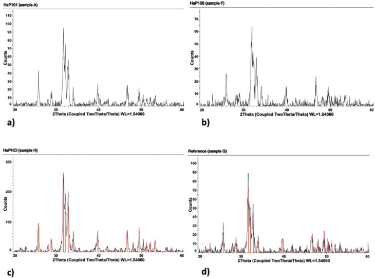
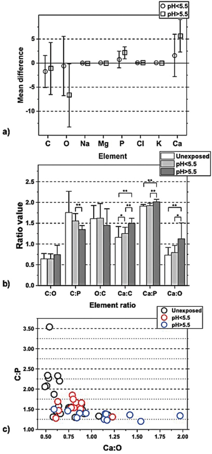
A number of fifteen human premolars were used. The enamel of the coronal half in six of the teeth, were exposed by H2O2. Nine teeth were prepared to enamel powder. The enamel was exposed to 3.3% H2O2, at six different pH-values (pH range 4.5 - 7.0). Analyses of the topography of enamel performed by scanning electron microscope (SEM) and the chemical composition of enamel by X-ray microanalysis (XRMA). X-ray powder diffraction (XRD) analysed the crystallinity in enamel powder.
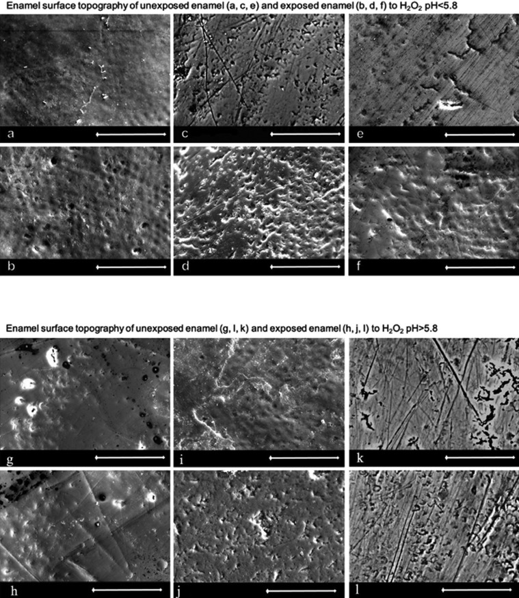
The exposure to H2O2 at pH<5.5 resulted in a rougher topography of the enamel, according to the SEM studies. The XRMA analysis revealed a increase in the ratio of Ca:C. Exposure to H2O2 at pH>5.5 resulted in a decrease of O in the exposed enamel, and changes in C:P, Ca:C, Ca:P and Ca:O were observed. The H2O22 did not affect the unit cell parameters, but the signal-to-noise level was increased for slightly acidic or neutral solutions. The unit cell parameters decreased in the acidic solutions.
The exposure to H2O2 at varying pH values affect the enamel with two different mechanisms. One effect is the oxidation of the organic or bioorganic matter in the hydroxyapatite matrix, due to the use of 3.3% H2O2. The other effect is due to the current pH of the H2O2, since the structure of the hydroxyapatite starts to erode when the pH<5.5. Dental Enamel, Tooth Bleaching Agents, Hydrogen Peroxide, Scanning Electron Microscopy, X-ray diffraction.
本研究展示了不同pH值的3.3%过氧化氢对牙釉质的影响。
使用了15颗人类前磨牙。其中6颗牙齿冠部一半的牙釉质用过氧化氢处理。另外9颗牙齿制备成牙釉质粉末。将牙釉质暴露于6种不同pH值(pH范围4.5 - 7.0)的3.3%过氧化氢中。通过扫描电子显微镜(SEM)分析牙釉质的表面形貌,并用X射线微分析(XRMA)分析牙釉质的化学成分。用X射线粉末衍射(XRD)分析牙釉质粉末的结晶度。
根据SEM研究,pH<5.5时暴露于过氧化氢会导致牙釉质表面形貌更粗糙。XRMA分析显示钙与碳的比例增加。pH>5.5时暴露于过氧化氢会导致暴露牙釉质中的氧减少,并且观察到碳与磷、钙与碳、钙与磷以及钙与氧的变化。过氧化氢并未影响晶胞参数,但对于微酸性或中性溶液,信噪比增加。酸性溶液中晶胞参数减小。
在不同pH值下暴露于过氧化氢会通过两种不同机制影响牙釉质。一种影响是由于使用3.3%过氧化氢导致羟基磷灰石基质中的有机或生物有机物质氧化。另一种影响是由于过氧化氢当前的pH值,因为当pH<5.5时羟基磷灰石结构开始侵蚀。牙釉质、牙齿漂白剂、过氧化氢、扫描电子显微镜、X射线衍射。