Zubizarreta-Macho Álvaro, Ferreiroa Alberto, Agustín-Panadero Rubén, Rico-Romano Cristina, Lobo-Galindo Ana-Belén, Mena-Álvarez Jesús
DDS, PhD. Associate professor. Department of Endodontics. Faculty of Health Sciences. Alfonso X el Sabio University. Madrid. Spain.
DDS, PhD. Associate professor. Department of Dental Prosthesis. Faculty of Dentistry. Complutense University. Madrid. Spain.
J Clin Exp Dent. 2019 Jun 1;11(6):e570-e576. doi: 10.4317/jced.55840. eCollection 2019 Jun.
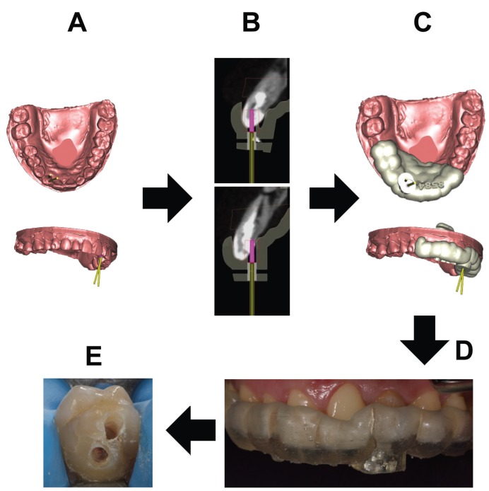
The complex anatomy of dens invaginatus makes access cavity to root canal system difficult, which has an impact on the prognosis of these teeth. A novel technique, based on new technologies, is proposed to make access cavity conservative and guided with minimal dental structure lost.
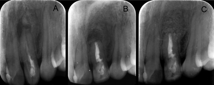
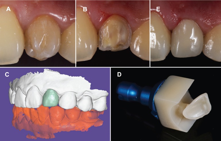
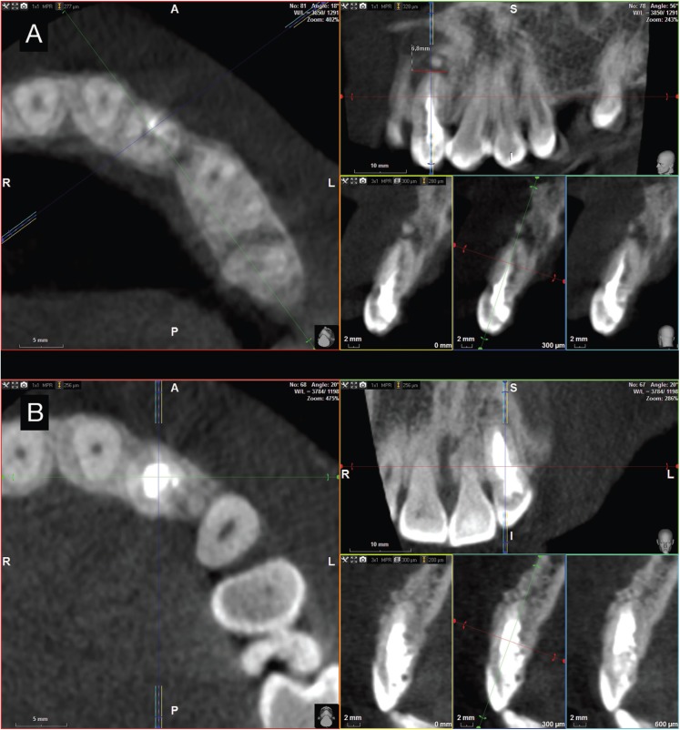
This case report shows the root canal retreatment and the endodontic surgery of a dens invaginatus type II in a left lateral upper incisor previously treated which was affected by a chronic apical abscess and an apical fracture. A Cone Beam Computed Tomography was performed to better diagnosis the dental anatomy. An intraoral scan was performed to get a digital 3D model. A computer-guided implant planning software was used to plan the access cavity and design the splint guided. Finally, the clinical crown was restored by a resin nanoceramic veneer made by a chairside system made up of an intraoral scanning unit and a grinding unit. Last, the authors carried through the endodontic surgery to extract the apical fractured fragment.
Follow-up appointments at 6, 12 and 18 months showed a radiographic reduction of the periapical lesion and absence of clinical signs.
The splint guide allowed a guided and conservative access cavity to root canal system. It facilitates the root canal retreatment and improves the prognosis of the teeth with dental malformations. CAD-CAM, Cone-Beam Computed Tomography, dens in dente, dens invaginatus, dental pulp cavity, endodontics.
牙内陷的复杂解剖结构使进入根管系统的预备洞形困难,这对这些牙齿的预后有影响。基于新技术提出了一种新颖的技术,以使预备洞形保守,并在牙体结构损失最小的情况下进行引导。
本病例报告展示了一例先前治疗过的左上侧切牙II型牙内陷的根管再治疗和牙髓外科手术,该牙受慢性根尖脓肿和根尖骨折影响。进行锥形束计算机断层扫描以更好地诊断牙齿解剖结构。进行口内扫描以获得数字3D模型。使用计算机引导的种植体规划软件来规划进入洞形并设计引导夹板。最后,通过由口内扫描单元和研磨单元组成的椅旁系统制作的树脂纳米陶瓷贴面修复临床冠。最后,作者进行牙髓外科手术取出根尖骨折碎片。
6个月、12个月和18个月的随访显示根尖周病变在影像学上缩小且无临床症状。
引导夹板允许对根管系统进行引导性和保守性的进入洞形预备。它有助于根管再治疗并改善牙畸形牙齿的预后。计算机辅助设计与制造、锥形束计算机断层扫描、牙中牙、牙内陷、牙髓腔、牙髓病学