Faculty of Veterinary Medicine, Universiti Putra Malaysia, Universiti Putra Malaysia, Serdang, 43400 Selangor, Malaysia.
Department of Family Medicine, School of Medical Sciences, USM Health Campus, 16150 Kubang Kerian, Kelantan, Malaysia.
Biomed Res Int. 2019 Jul 3;2019:6979585. doi: 10.1155/2019/6979585. eCollection 2019.
The objective of the study is to evaluate the chondroprotective activity of (Channa) and glucosamine sulphate (glucosamine) on histomorphometric examinations, serum biomarker, and inflammatory mediators in experimental osteoarthritis (OA) rabbit model.
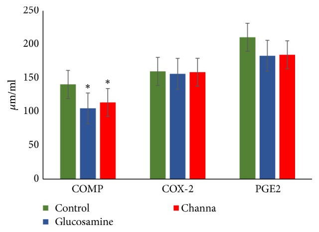
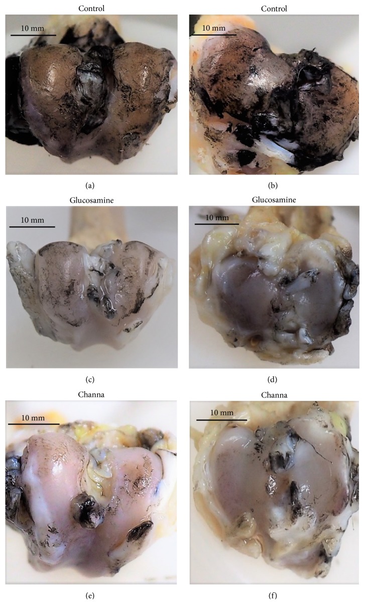
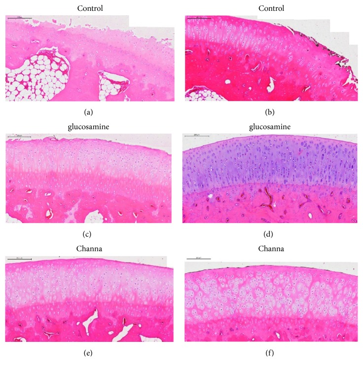
Anterior cruciate ligament transection (ACLT) was performed to induce OA in thirty-three male New Zealand white rabbits and were randomly divided into three groups: Channa, glucosamine, and control group. The control group received drinking water and the Channa and glucosamine groups were orally administered with 51.4 mg/kg of Channa extract and 77.5 mg/kg of glucosamine sulphate in drinking water, respectively, for eight weeks and then sacrificed. The articular cartilage was evaluated macroscopically and histologically using semiquantitative and quantitative methods. Serum cartilage oligomeric matric protein (COMP), cyclooxygenase 2 (COX-2) enzyme, and prostaglandin E (PGE) were also determined.
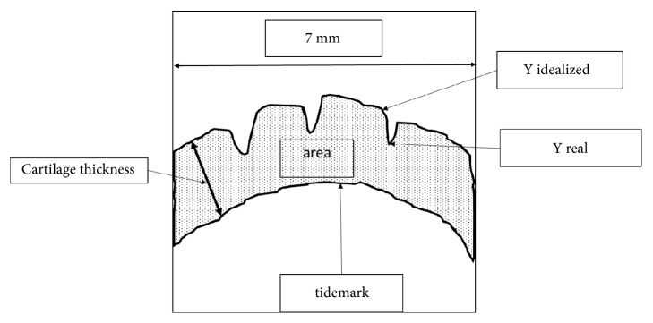
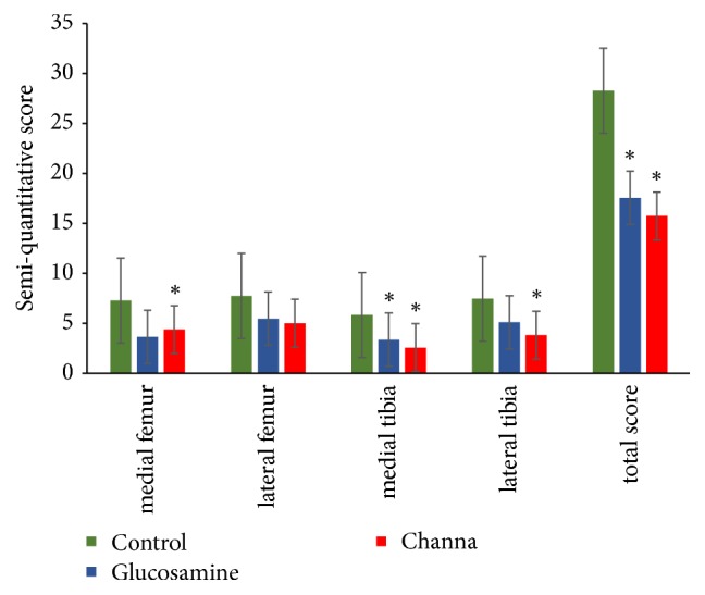
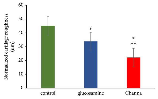
Macroscopic analysis revealed that Channa group have a significantly lower severity grade of total macroscopic score compared to the control (p < 0.001) and glucosamine (p < 0.05) groups. Semiquantitative histology scoring showed that both Channa and glucosamine groups had lower severity grading of total histology score compared to the control group (p < 0.001). In comparison with the control, Channa group had lower histopathological changes in three compartments of the joint compared to glucosamine group which had lower histological scoring in two compartments only. The cartilage thickness, area, and roughness of both Channa (p < 0.05) and glucosamine (p < 0.05) groups were superior compared to the control group. However, the Channa group demonstrated significantly less cartilage roughness compared to the glucosamine group (p < 0.05). Serum COMP levels were lower in both Channa (p < 0.05) and glucosamine (p < 0.05) groups compared to the control group.
Both oral administration of Channa extract and glucosamine exhibited chondroprotective action on an ACLT OA-induced rabbit model. However, Channa was superior to glucosamine in maintaining the structure of the cartilage.
本研究旨在通过组织形态计量学检查、血清生物标志物和炎症介质评估 (查那) 和硫酸葡聚糖胺(葡聚糖胺)对实验性骨关节炎(OA)兔模型的软骨保护活性。
通过前交叉韧带切断术(ACLT)诱导 33 只雄性新西兰白兔发生 OA,并将其随机分为三组:查那组、葡聚糖胺组和对照组。对照组给予饮用水,查那组和葡聚糖胺组分别给予 51.4mg/kg 的查那提取物和 77.5mg/kg 的硫酸葡聚糖胺口服,连续 8 周后处死。采用半定量和定量方法评估关节软骨的大体和组织学表现。还测定了血清软骨寡聚基质蛋白(COMP)、环氧化酶 2(COX-2)酶和前列腺素 E(PGE)。
大体分析显示,与对照组(p<0.001)和葡聚糖胺组(p<0.05)相比,查那组的总大体评分严重程度分级明显较低。半定量组织学评分显示,与对照组相比,查那组和葡聚糖胺组的总组织学评分严重程度分级均较低(p<0.001)。与对照组相比,查那组的关节三个部位的组织病理学变化较小,而葡聚糖胺组仅两个部位的组织学评分较低。与对照组相比,查那组和葡聚糖胺组的软骨厚度、面积和粗糙度均有改善(p<0.05)。然而,查那组的软骨粗糙度明显低于葡聚糖胺组(p<0.05)。与对照组相比,查那组(p<0.05)和葡聚糖胺组(p<0.05)的血清 COMP 水平均较低。
查那提取物和葡聚糖胺的口服给药均对 ACLT 诱导的 OA 兔模型表现出软骨保护作用。然而,查那在维持软骨结构方面优于葡聚糖胺。