Univ Rennes, Inria, CNRS, INSERM, IRISA, Empenn ERL U-1228, F-35000 Rennes, France; CHU Rennes, Radiology Department, F-35033 Rennes, France.
Univ Rennes, Inria, CNRS, INSERM, IRISA, Empenn ERL U-1228, F-35000 Rennes, France.
Neuroimage Clin. 2019;24:101939. doi: 10.1016/j.nicl.2019.101939. Epub 2019 Jul 16.
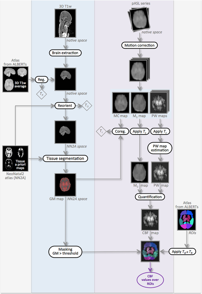
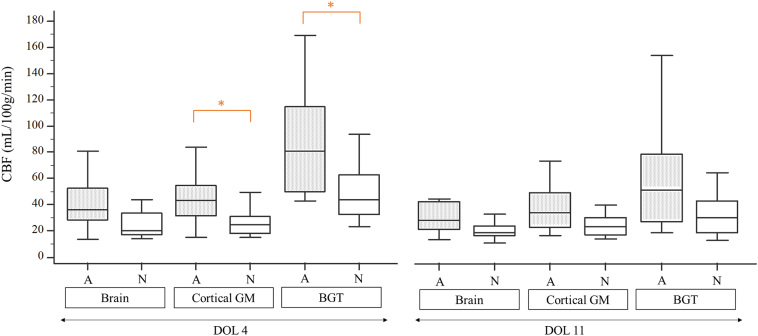
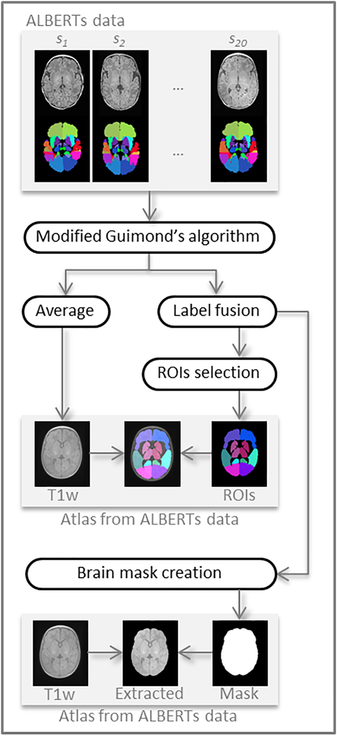
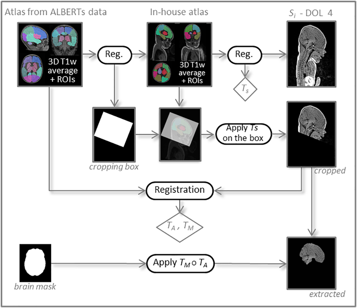
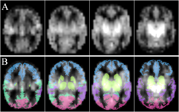
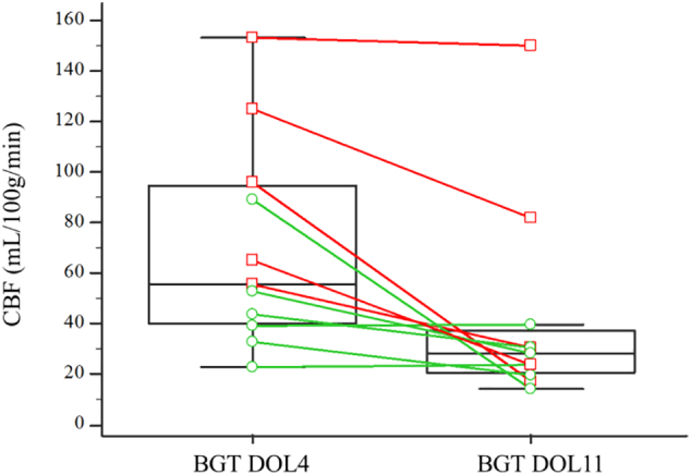
The primary objective of this study was to evaluate changes in cerebral blood flow (CBF) using arterial spin labeling MRI between day 4 of life (DOL4) and day 11 of life (DOL11) in neonates with hypoxic-ischemic encephalopathy (HIE) treated with hypothermia. The secondary objectives were to compare CBF values between the different regions of interest (ROIs) and between infants with ischemic lesions on MRI and infants with normal MRI findings. We prospectively included all consecutive neonates with HIE admitted to the neonatal intensive care unit of our institution who were eligible for therapeutic hypothermia. Each neonate systematically underwent two MRI examinations as close as possible to day 4 (early MRI) and day 11 (late MRI) of life. A custom processing pipeline of morphological and perfusion imaging data adapted to neonates was developed to perform automated ROI analysis. Twenty-eight neonates were included in the study between April 2015 and December 2017. There were 16 boys and 12 girls. Statistical analysis was finally performed on 37 MRIs, 17 early MRIs and 20 late MRIs. Eleven neonates had both early and late MRIs of good quality available. Eight out of 17 neonates (47%) had an abnormal on late MRI as performed and 7/20 neonates (35%) had an abnormal late MRI. CBF values in the basal ganglia and thalami (BGT) and temporal lobes were significantly higher on DOL4 than on DOL11. There were no significant differences between DOL4 and DOL11 for the other ROIs. CBF values were significantly higher in the BGT vs. the cortical GM, on both DOL4 and DOL11. On DOL4, the CBF was significantly higher in the cortical GM, the BGT, and the frontal and parietal lobes in subjects with an abnormal MRI compared to those with a normal MRI. On DOL11, CBF values in each ROI were not significantly different between the normal MRI group and the abnormal MRI group, except for the temporal lobes. This article proposes an innovative processing pipeline for morphological and ASL data suited to neonates that enable automated segmentation to obtain CBF values over ROIs. We evaluate CBF on two successive scans within the first 15 days of life in the same subjects. ASL imaging in asphyxiated neonates seems more relevant when used relatively early, in the first days of life. The correlation of intra-subject changes in cerebral perfusion between early and late MRI with neurodevelopmental outcome warrants investigation in a larger cohort, to determine whether the CBF pattern change can provide prognostic information beyond that provided by visible structural abnormalities on conventional MRI.
本研究的主要目的是评估在接受低温治疗的患有缺氧缺血性脑病(HIE)的新生儿中,在第 4 天(DOL4)和第 11 天(DOL11)之间使用动脉自旋标记 MRI 评估脑血流(CBF)的变化。次要目标是比较不同感兴趣区域(ROI)之间以及 MRI 上有缺血性病变的婴儿与 MRI 正常发现的婴儿之间的 CBF 值。我们前瞻性地纳入了所有符合条件接受治疗性低温治疗的患有 HIE 的连续入院新生儿。每个新生儿都系统地进行了两次 MRI 检查,尽可能接近生命的第 4 天(早期 MRI)和第 11 天(晚期 MRI)。开发了一种适用于新生儿的形态和灌注成像数据的定制处理管道,以进行自动 ROI 分析。2015 年 4 月至 2017 年 12 月期间,共有 28 名新生儿纳入研究。其中 16 名男孩和 12 名女孩。最终对 37 次 MRI、17 次早期 MRI 和 20 次晚期 MRI 进行了统计分析。11 名新生儿有质量良好的早期和晚期 MRI 可用。17 名新生儿中有 8 名(47%)晚期 MRI 异常,20 名新生儿中有 7 名(35%)晚期 MRI 异常。DOL4 时基底节和丘脑(BGT)和颞叶的 CBF 值明显高于 DOL11。其他 ROI 之间 DOL4 和 DOL11 之间没有显着差异。DOL4 和 DOL11 时,BGT 与皮质 GM 的 CBF 值均较高。在 DOL4 时,与 MRI 正常的受试者相比,MRI 异常的受试者的皮质 GM、BGT 和额叶和顶叶的 CBF 明显更高。在 DOL11 时,除了颞叶,正常 MRI 组和异常 MRI 组之间各 ROI 的 CBF 值没有显着差异。本文提出了一种适用于新生儿的创新形态和 ASL 数据处理管道,可实现自动分割以获得 ROI 上的 CBF 值。我们在同一受试者的生命最初 15 天内评估了两次连续扫描的 CBF。在生命的最初几天,在窒息的新生儿中使用 ASL 成像似乎更为相关。在较大的队列中,需要研究亚组间早期和晚期 MRI 之间脑灌注变化与神经发育结局之间的相关性,以确定 CBF 模式变化是否可以提供比常规 MRI 上可见的结构性异常提供的预后信息更多的预后信息。