Tu Liyun, Porras Antonio R, Oh Albert, Lepore Natasha, Mastromanolis Manuel, Tsering Deki, Paniagua Beatriz, Enquobahrie Andinet, Keating Robert, Rogers Gary F, Linguraru Marius George
Sheikh Zayed Institute for Pediatric Surgical Innovation, Children's National Health System, Washington DC, USA.
Division of Plastic and Reconstructive Surgery, Children's National Health System, Washington DC, USA.
Proc SPIE Int Soc Opt Eng. 2018 Feb;10575. doi: 10.1117/12.2295374. Epub 2018 Feb 27.
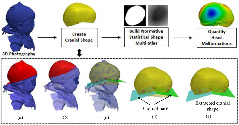
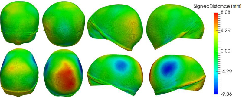
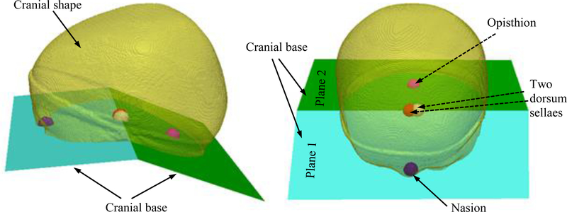
The evaluation of cranial malformations plays an essential role both in the early diagnosis and in the decision to perform surgical treatment for craniosynostosis. In clinical practice, both cranial shape and suture fusion are evaluated using CT images, which involve the use of harmful radiation on children. Three-dimensional (3D) photography offers non-invasive, radiation-free, and anesthetic-free evaluation of craniofacial morphology. The aim of this study is to develop an automated framework to objectively quantify cranial malformations in patients with craniosynostosis from 3D photography. We propose a new method that automatically extracts the cranial shape by identifying a set of landmarks from a 3D photograph. Specifically, it registers the 3D photograph of a patient to a reference template in which the position of the landmarks is known. Then, the method finds the closest cranial shape to that of the patient from a normative statistical shape multi-atlas built from 3D photographs of healthy cases, and uses it to quantify objectively cranial malformations. We calculated the cranial malformations on 17 craniosynostosis patients and we compared them with the malformations of the normative population used to build the multi-atlas. The average malformations of the craniosynostosis cases were 2.68 ± 0.75 mm, which is significantly higher (p<0.001) than the average malformations of 1.70 ± 0.41 mm obtained from the normative cases. Our approach can support the quantitative assessment of surgical procedures for cranial vault reconstruction without exposing pediatric patients to harmful radiation.
颅骨畸形的评估在早期诊断以及决定是否对颅缝早闭进行手术治疗方面都起着至关重要的作用。在临床实践中,颅骨形状和缝线融合情况都是通过CT图像来评估的,而这会让儿童受到有害辐射。三维(3D)摄影提供了一种非侵入性、无辐射且无需麻醉的颅面形态评估方法。本研究的目的是开发一个自动化框架,以便从3D摄影中客观量化颅缝早闭患者的颅骨畸形。我们提出了一种新方法,通过从3D照片中识别一组地标来自动提取颅骨形状。具体而言,它将患者的3D照片与一个已知地标位置的参考模板进行配准。然后,该方法从由健康病例的3D照片构建的规范统计形状多图谱中找到与患者颅骨形状最接近的形状,并使用它来客观量化颅骨畸形。我们计算了17例颅缝早闭患者的颅骨畸形情况,并将其与用于构建多图谱的正常人群的畸形情况进行了比较。颅缝早闭病例的平均畸形为2.68±0.75毫米,显著高于(p<0.001)从正常病例中获得的1.70±0.41毫米的平均畸形。我们的方法可以支持对颅盖重建手术进行定量评估,而无需让儿科患者暴露于有害辐射之下。