Reinert Michael, Piffaretti Deborah, Wilzbach Marco, Hauger Christian, Guckler Roland, Marchi Francesco, D'Angelo Maria Luisa
Laboratory for Biomedical Neurosciences, Neurocenter of Southern Switzerland, Ente Ospedaliero Cantonale, Torricella-Taverne, Switzerland.
Department of Neurosurgery, Neurocenter of Southern Switzerland, Ente Ospedaliero Cantonale, Lugano, Switzerland.
Front Surg. 2019 Jul 16;6:41. doi: 10.3389/fsurg.2019.00041. eCollection 2019.
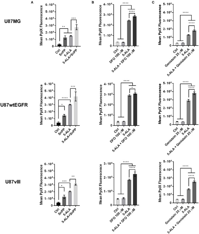
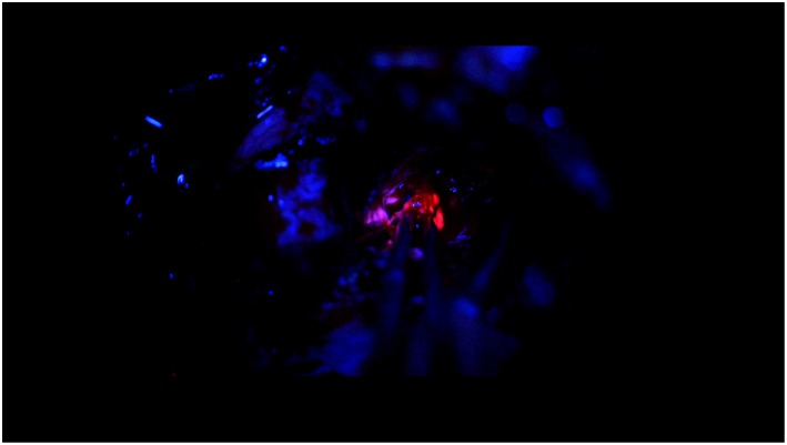
5-Aminolevulinic acid (5-ALA) induced fluorescence to augment surgical resection for high grade glioma has become a standard of care. Protoporphyrin IX (PpIX) visibility is however subject to the variability of the single tumor expression and to the interobserver interpretation. We therefore hypothesized that in different glioma cell lines with variable 5-ALA induced fluorescence, the signal can be pharmacologically increased. We therefore analyzed in three different GBM cell lines, with different expression of epidermal growth factor receptor (EGFR), the variability of 5-ALA induced PpIX fluorescence after the pharmacological blockade at different steps of PpIX breakdown and influencing the outbound transport of PpIX. Using flow cytometry, fluorescence microplate reader, and confocal microscopy the PpIX fluorescence was analyzed after exposure to tin protoporphyrin IX (SnPP), deferoxamine (DFO), and genistein. We furthermore constructed a microscope (Qp9-microscope) being able to measure quantitatively the concentration of PpIX. These values were compared with the extraction of PpIX in tumor biopsy taken during the GBM surgery. Although all three cell lines showed an increase to 5-ALA induced fluorescence their baseline activity was different. Treatment with either SnPP, DFO and genistein was able to increase 5-ALA induced fluorescence. Qp9-microscopy of tumor sample produced a color coded PpIX concentration map which was overlaid on the tumor image. The PpIX extraction from tumor sample analyzed using the plate reader gave lower values of the concentration, as compared to the expected values of the Qp9-microscope, however still in the same decimal range of μg/mL. This may be due to homogenization of the values during extraction and cell disaggregation. In conclusion pharmacological augmentation in GBM cell lines of PpIX signal is possible. A quantitative PpIX map for surgery is feasible and may help refine surgical excision. Further correlations of tumor tissue samples and Qp9-microscopy is needed, prior to develop an intraoperative surgical adjunct to the already existing 5-ALA induced surgery.
5-氨基乙酰丙酸(5-ALA)诱导荧光以增强高级别胶质瘤的手术切除已成为一种标准治疗方法。然而,原卟啉IX(PpIX)的可见性受单个肿瘤表达的变异性和观察者间解释的影响。因此,我们假设在具有可变5-ALA诱导荧光的不同胶质瘤细胞系中,该信号可通过药理学方法增强。因此,我们在三种不同的胶质母细胞瘤(GBM)细胞系中进行分析,这些细胞系表皮生长因子受体(EGFR)表达不同,在PpIX分解的不同步骤进行药理学阻断并影响PpIX的外向转运后,分析5-ALA诱导的PpIX荧光的变异性。使用流式细胞仪、荧光微孔板读数器和共聚焦显微镜,在暴露于锡原卟啉IX(SnPP)、去铁胺(DFO)和染料木黄酮后分析PpIX荧光。我们还构建了一台能够定量测量PpIX浓度的显微镜(Qp9显微镜)。将这些值与GBM手术期间获取的肿瘤活检中PpIX的提取量进行比较。尽管所有三种细胞系对5-ALA诱导的荧光均有增加,但其基线活性不同。用SnPP、DFO和染料木黄酮治疗均能增加5-ALA诱导的荧光。肿瘤样本的Qp9显微镜检查产生了一个颜色编码的PpIX浓度图,该图叠加在肿瘤图像上。与Qp9显微镜的预期值相比,使用酶标仪分析肿瘤样本中PpIX的提取量得到的浓度值较低,但仍在相同的μg/mL数量级范围内。这可能是由于提取过程中和细胞解离过程中值的均一化所致。总之,在GBM细胞系中通过药理学方法增强PpIX信号是可能的。用于手术的定量PpIX图是可行的,可能有助于完善手术切除。在开发针对现有5-ALA诱导手术的术中手术辅助设备之前,需要进一步将肿瘤组织样本与Qp9显微镜检查进行相关性研究。