Akhter Asad S, El Tecle Najib, Alexopoulos Georgios, Espinoza Gabriela, Coppens Jeroen
Neurosurgery, Ohio State University, Wexner Medical Center, Colombus, USA.
Neurosurgery, St. Louis University Hospital, St. Louis, USA.
Cureus. 2019 Jun 4;11(6):e4823. doi: 10.7759/cureus.4823.
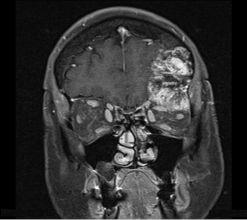
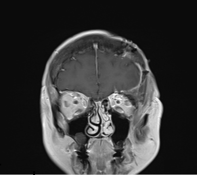
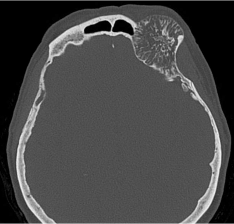
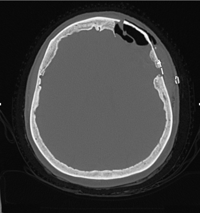
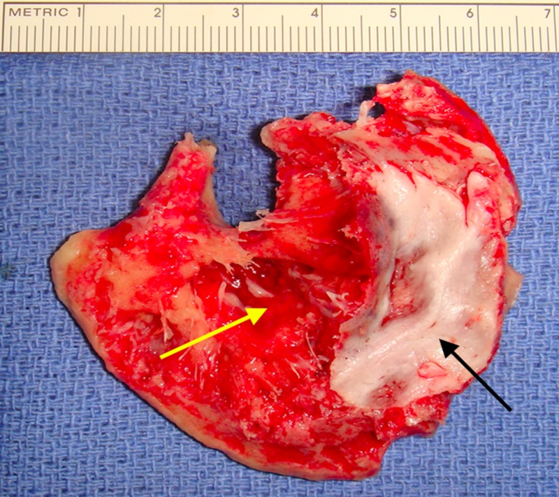
Primary intraosseous cavernous hemangiomas are rare skull lesions that are not typically known to involve the orbital bones or the dura. We describe a rare case of a fronto-orbital bone cavernous hemangioma with extension into the dura. A 68-year-old female presented with a one-year history of diplopia with discomfort around her left orbit. Magnetic resonance images demonstrated a mass in the left frontal skull extending into the orbital rim. The patient underwent a craniotomy for tumor resection. Dural invasion was found intraoperatively. Gross total resection and reconstruction were achieved. On the postoperative follow-up, the patient was asymptomatic. Primary calvarial intraosseous cavernous hemangiomas are most commonly located in the frontal and parietal bones. These lesions typically involve only the outer table of the skull. In lesions involving the orbit and dura, excision with cranioplasty can provide symptomatic relief with good cosmetic outcomes.
原发性骨内海绵状血管瘤是罕见的颅骨病变,通常不会累及眶骨或硬脑膜。我们描述了一例罕见的额眶骨海绵状血管瘤并累及硬脑膜的病例。一名68岁女性,有一年复视病史,伴有左眼眶周围不适。磁共振成像显示左额颅骨有一肿块延伸至眶缘。患者接受了开颅肿瘤切除术。术中发现硬脑膜受侵。实现了肿瘤全切及重建。术后随访时,患者无症状。原发性颅骨内海绵状血管瘤最常见于额骨和顶骨。这些病变通常仅累及颅骨外板。对于累及眼眶和硬脑膜的病变,行颅骨成形术切除可缓解症状并获得良好的美容效果。