National Innovation and Attracting Talents "111" base, Key Laboratory of Biorheological Science and Technology, Ministry of Education, College of Bioengineering, Chongqing University, Chongqing, China.
Hubei Engineering Technology Research Center of TCM Processing, College of Pharmacy, Hubei University of Chinese Medicine, Wuhan, China.
Cell Prolif. 2019 Sep;52(5):e12666. doi: 10.1111/cpr.12666. Epub 2019 Aug 12.
Cartilaginous tissue degradation occurs because of the lack of survival of chondrocytes. Here, we ascertained whether bakuchiol (BAK) has the capability of activating chondrocyte proliferation.
The effect of BAK on the proliferation of rat chondrocytes at a concentration of 10 and 20 µmol/L was investigated. The molecular mechanisms involving target binding and signalling pathways were elucidated by RNA-sequencing, qPCR, molecular docking and Western blotting. Matrigel mixed with bakuchiol was implanted locally into rat knee articular cartilage defects to verify the activation of chondrocytes due to bakuchiol in vivo.
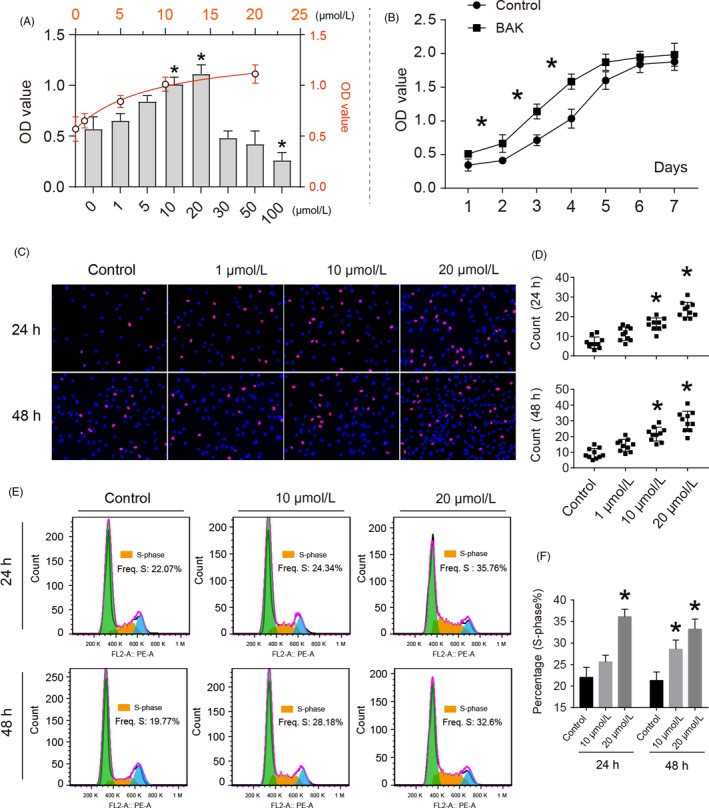
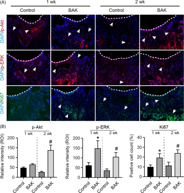
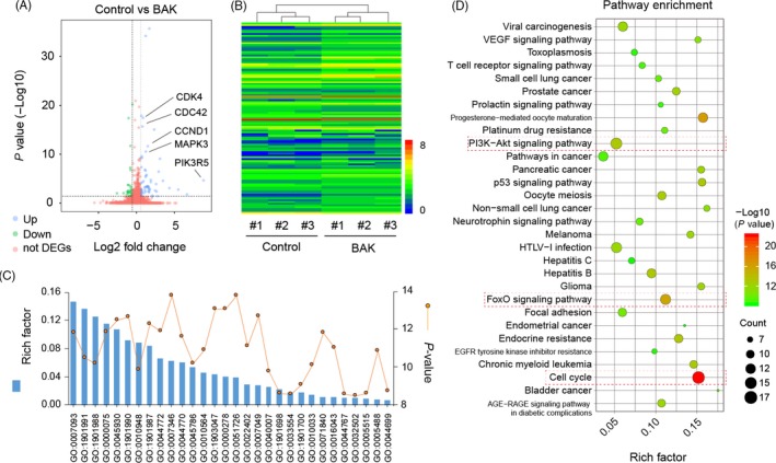
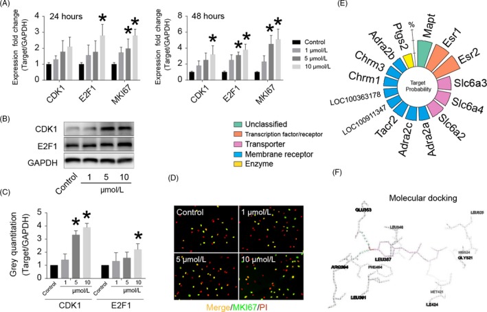
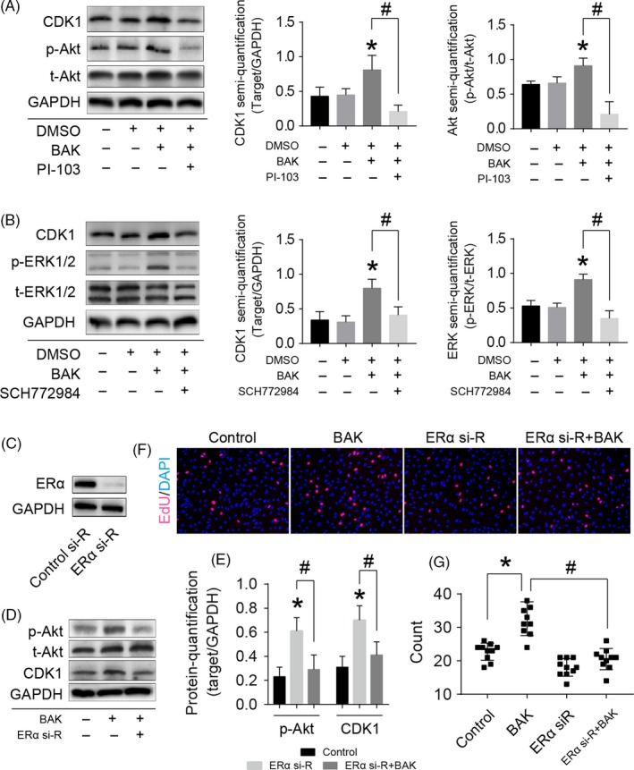
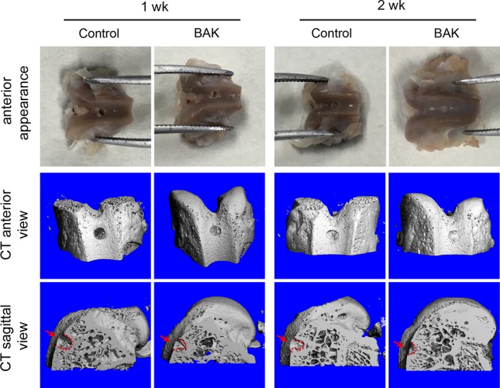
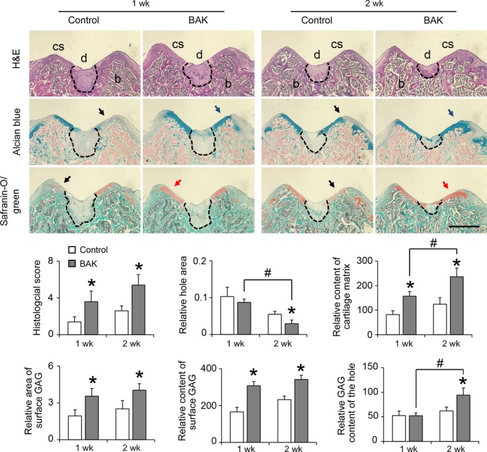
Bakuchiol implantation resulted in the activation of rat chondrocyte proliferation in a dose-dependent manner. RNA-sequencing revealed 107 differentially expressed genes (DEGs) with 75 that were up-regulated and 32 that were down-regulated, indicating increased activation of the PI3K-Akt and cell cycle pathways. Activation of the phosphorylation of Akt, ERK1/2 and their inhibitors blocked the proliferative effect of bakuchiol treatment, confirming its direct involvement in these signal transduction pathways. Molecular docking and siRNA silencing revealed that estrogen receptor-α (ERα) was the target of bakuchiol in terms of its cell proliferative effect via PI3K activation. Two weeks after implantation of bakuchiol, the appearance and physiological structure of the articular cartilage was more integrated with abundant chondrocytes and cartilage matrix compared to that of the control.
Bakuchiol demonstrated significant bioactivity towards chondrocyte proliferation via the PI3K-Akt and ERK1/2 pathways mediated by estrogen receptor activation and exhibited enhanced promotion of the remodelling of injured cartilage.
软骨组织降解是由于软骨细胞缺乏生存能力所致。在这里,我们确定补骨脂素(BAK)是否具有激活软骨细胞增殖的能力。
研究了 BAK 在浓度为 10 和 20 μmol/L 时对大鼠软骨细胞增殖的影响。通过 RNA 测序、qPCR、分子对接和 Western blot 阐明了涉及靶标结合和信号通路的分子机制。将含有补骨脂素的 Matrigel 局部植入大鼠膝关节关节软骨缺损处,以验证补骨脂素在体内激活软骨细胞的作用。
补骨脂素植入以剂量依赖的方式导致大鼠软骨细胞增殖的激活。RNA 测序显示 107 个差异表达基因(DEGs),其中 75 个上调,32 个下调,表明 PI3K-Akt 和细胞周期途径的激活增加。Akt、ERK1/2 的磷酸化激活及其抑制剂的激活阻断了补骨脂素处理的增殖作用,证实了其直接参与这些信号转导途径。分子对接和 siRNA 沉默表明,雌激素受体-α(ERα)是补骨脂素通过激活 PI3K 发挥其细胞增殖作用的靶标。植入补骨脂素两周后,与对照组相比,关节软骨的外观和生理结构更加完整,有丰富的软骨细胞和软骨基质。
补骨脂素通过雌激素受体激活介导的 PI3K-Akt 和 ERK1/2 途径对软骨细胞增殖表现出显著的生物活性,并表现出增强对受损软骨重塑的促进作用。